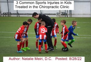
Sports-related injuries in kids and adolescents (teenagers) are frequent and can be helped in the chiropractic clinic. Over 7 million teenagers are involved in high school sports.  Over 20 million children between the ages of 8 and 16 years are involved in non-school community athletic programs.  Many participate in unstructured, relatively unsupervised recreation. And almost all middle and junior high schools have sports programs.


Kids and teenagers are young athletes with anatomic and physiologic uniqueness in how they participate in sports activities. As always, prevention is still a major goal even for a child athlete.  When injured, a proper diagnosis, management and rehabilitation is critical for children and adolescents.


While children generally participate in a sport(s) activity for fun, there are other important considerations.  Firstly, children attain self-confidence through participation. Secondly, it develops their socializing skills and avoids boredom. Thirdly, it may help them to learn about setting goals.  Fifthly, physical participation in sports lays the groundwork for a healthy lifestyle. In other words, exercise habits carry over into adulthood. In fact, studies have found the percentage of body fat of adults who exercised as adolescents is lower.  Lastly, exercise may affect physical growth. So minimum amounts of exercise may stimulate growth while excessive exercise may retard growth. 

3 Common Sports Injuries in Kids Treated in the Chiropractic Clinic


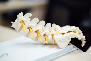
1.  Spondylolisthesis, Spondylolysis and Low Back Pain


Spondylolysis occurs in approximately 6 % of the population. Spondylolysis is more common in female gymnasts, college football linemen, weight lifters, and rowers. Moreover, risk is also increased during growth spurts.


Spondylolysis is a stress fracture through the pars interarticularis of the lumbar vertebrae.  The pars is a small segment of bone that joins the facet joints (joint connects two or more bones) in the back of the spine. A defect in this portion of the spine leads to this condition called spondylolysis as well.


So Spondylolysis is due either from a stress fracture of the pars interarticularis or an elongated (lengthened) pars. The elongated pars is a result from micro (small) fractures that heal with an elongated pars. And a microfracture could be caused by trauma, like getting hit by something or a fall. Also, a stress fracture may be due to repetitive micro trauma such as hyperextension (extensive arching of the back) in sports activities.


Spondylolisthesis is a condition in which a vertebra (bone) in the spine moves forward out of the proper position onto the bone below it. There are several types of spondylolisthesis. However, the most common is isthmic (as a result of spondylolysis), occurring in the young. Although congenital (present from birth) types and destructive (e.g., tuberculosis,cancer) types are possible, they are rare.


In conclusion, there is a good response to chiropractic manipulative treatment for low back pain due to spondylolithesis (grades 1 or 2) resulting from a spondylysis. 


Spondylolisthesis is primarily a radiographic (x-ray) diagnosis.


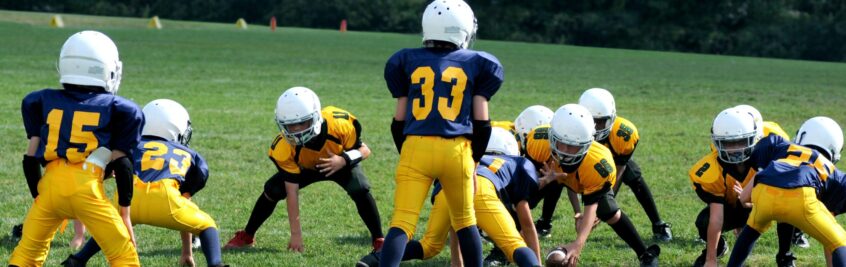
2. Neck Injury/ “Burner/Stinger”


Lateral flexion neck (moving your head toward one of your shoulders) injuries often result in a “burner” or “stinger” seen commonly in sports with kids and adolescents. This is a common injury in sports and often recur leading to further disability. It is a common injury in football hockey, wrestling, lacrosse, and diving. 


 “Burner” or “stinger” are the names given to injury of the brachial plexus (network of nerves in the shoulder that carries movement and sensory signals from the spinal cord to the arms and hands) or nerve roots (initial segment of a nerve leaving spinal cord). In general, the injury is a lateral flexion of the head away from the involved side with accompanying shoulder distraction (depression) on the involved side.


Kids may have a sudden onset of burning pain and/or numbness along the lateral (side) arm with associated arm weakness following a lateral flexion injury of the neck (e.g., lateral “whiplash”). The symptoms of burning usually last only a couple of minutes. So while burners/stingers are typically transient (lasting a short time), kids may continue to have neck and arm pain or weakness afterwards. 


Radiographic (x-ray) evaluation is recommended to determine if any instability is present. If instability is present, it is necessary to consider restrictions in sports (e.g., football) where head and neck trauma is common to prevent further damage.


3) Osgood-Schlatter’s Apophyseal Injury and Knee/Leg Pain


Apophyseal injuries are stresses to the growth plate in adolescent athletes. Apophyseal injuries are almost always due to sports activity in kids. Osgood-Schlatter’s apophysitis at the tibial tubercle (secondary ossification center on the top of the shin bone) is commonly seen in running/jumping sports, in hockey with boys and gymnastics and figure skating for girls. Symptoms of pain and swelling are present over the tibial tuberosity below the knee joint, where the patellar tendon attaches to the top of the shinbone (tibia). There may also be inflammation of the patellar tendon, which stretches over the patella (kneecap).


First, activity modification (reduction of intensity or frequency) and icing is recommended.  Second, Chiropractic manipulative therapy (adjustments) should include a biomechanical assessment of the whole kinematic chain including all the joints and the muscles of the lower limb, the spine and pelvic joints. Third, soft tissue manipulation (e.g. myofascial release, pressure point, post-isometric relaxation) should be done on these muscles/tendon: hamstring, quadriceps, iliotibial band,and Achilles tendon.  Fourth, stretching,and strengthening of the indicated muscles are important. Taping and a  brace may provide some limited symptom relief. Osgood-Schlatter’s Apophyseal Injury takes about 6- 9 months to heal in most cases.


Radiographs (x-rays) are generally used either to rule out a tumor (if suspected) and/or a full avulsion injury.


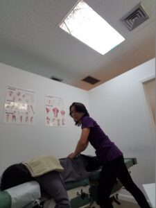
Safety of Chiropractic for Kids


The American Chiropractic Association cites a study done in 2014 confirming that chiropractic adjustments are overwhelmingly safe in infants and children.


In 2009, a survey found that there were about 68 million pediatric visits to chiropractors. The National Board of Chiropractic Examiners’ most recent practice analysis, issued in 2010, found that about 17 percent of chiropractic patients were under age 18 — approximately 7.7 percent aged five years or younger and some 9.4 percent between ages six and 17.


Children have the same joints that adults do, but not fully formed yet.  They are still developing and adjustments get the spine back in alignment. This keeps the body balanced and to prevent further problems. Also, Kids joints are still more cartilaginous than truly bony, so the adjustments have to be a little bit faster, but with less force. This is due to the increased flexibility within the joint and the smaller surface area targeted. 


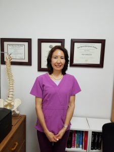
Dr. Natalie Meiri delivers fast adjustments that are low intensity (low force) and low amplitude (low displacement).  She also has helped babies and kids utilizing soft tissue techniques, cranial sacral therapy, and homeopathy. She has treated babies with ear infections, kids with ADHD, autism, scoliosis, sports injuries and various growing problems.  Contact her at 561-253-8984 for further information on 3 Common Sports Injuries in Kids Treated in the Chiropractic Clinic or to make an appointment.


This post is about Chiropractic Care of Shoulder Girdle Pain: Acromioclavicular Joint Injury. This injury can occur in any age group, but occurs mostly in 20-29 year olds.  You will have shoulder pain following a traumatic event.  Moreover, you may have had a fall onto the top of your shoulder or on an outstretched arm. It also occurs during a whiplash injury due to a car accident or a sports related injury.

If you have Acromioclavicular Joint Pain, Chiropractic can provide relief!


Acromioclavicular Joint Injury Symptoms and Signs

You will have local tenderness over the acromioclavicular joint. And you may experience painful decreased range of motion in the area.  Many of your activities will be limited including dressing and carrying and/or pulling objects.  Additionally, sleeping/lying on the injured shoulder side will be painful. Furthermore, in the case of a (grade III) sprain, obvious anatomical deformity and swelling will be apparent.  


Acromioclavicular Joint Anatomy


Firstly, your pectoral girdle (shoulder girdle), consists of your collar bone (clavicle) and your shoulder blade (scapula) which provides the bony link between your arm and your trunk.


Secondly, the acromioclavicular joint is formed by the cap of the shoulder blade (acromion of scapula) and the collar bone (clavicle). The outer end of the clavicle is held in alignment with the acromion by the acromioclavicular and coracoclavicular ligaments.


Thirdly, the acromioclavicular joint has a thin joint capsule lined with synovium (connective tissue that lines the inside of the joint capsule). A joint capsule, also called an articular capsule, is a bubble-like structure that surrounds joints such as the shoulder, elbow, wrist, hand, knee, foot, and ankle.


Fourthly, the acromioclavicular joint capsule is weak and is strengthened by the ligaments (acromioclavicular and coracoclavicular ligaments). These in turn are reinforced through attachments from the deltoid and trapezius muscles. Without these ligaments, the acromioclavicular joint capsule would not be strong enough to maintain the integrity of the joint.


Chiropractic Care of Shoulder Girdle Pain: Acromioclavicular Joint Injury Types I-III


An acromioclavicular joint separation or sprain is a traumatic separation or compression of the acromioclavicular joint.


Acromioclavicular joint separations are classified into three grades treated and managed in the chiropractic office:


First degree- Grade I- indicates some tearing of the acromioclavicular ligament, but no instability.


Second degree- Grade II- indicates rupture of the acromioclavicular ligament.


Third degree- Grade III involves tearing both of the acromioclavicular and the coracoclavicular ligaments. Both grades II and III are unstable (requires short period of immobilization in a sling). And type III injuries often take longer to heal (several weeks to months compared to Type I, II).

adjustment of patient's shoulder


Chiropractic Care of Shoulder Girdle Pain: Acromioclavicular Joint Injury Treatment


Chiropractic manipulative treatment/therapy to the spine and extremities (e.g. acromioclavicular joint, glenohumeral joint) are effective for acromioclavicular joint injuries. Our other therapies, such as soft tissue techniques, electric muscle stimulation, ice/ heat therapies and homeopathic consultations provide relief.

Indeed, acromioclavicular separations may need a short period of support with a shoulder sling (type II-III).  Similarly, rehabilitation exercise starts with mild isometrics (even while in a sling).  Next isotonic shoulder exercises with an emphasis on deltoid and upper trapezius exercises followed by rotator cuff, biceps and pectoral exercises are important. 

Through regular chiropractic visits, you can get pain relief and improve your health without drugs or surgery!


At Meiri Chiropractic we spend the time necessary to examine, diagnose and treat every neuromusculoskeletal condition and various ailments you have.  Chiropractic is a holistic and natural way to not only treat existing conditions, but to keep your body in its best working condition.  Meiri Chiropractic has been offering effective chiropractic care in Palm Beach county since 2006.  Many of our patient reviews note our excellence.  Call us today at 561-253-8984 to make an appointment or to find out more about Chiropractic Care of Shoulder Girdle Pain: Acromioclavicular Joint Injury.


This post is about Sciatica: A Chiropractic Center Can Provide Relief.  Symptoms include mid-buttock pain that radiates down the leg, most often unilaterally (one side). You will often experience an abnormal sensation such as numbness or parethesia (pins and needles). You may also have weakness in the leg effected.

If you have Sciatica, Chiropractic can provide relief! We’ve got some great information to share below.

What is Sciatica?


Firstly, the sciatic nerve is the largest nerve in the human body. Sciatic nerves originate from several nerves in your lower back. These nerves branch outward from the spine and then come together at each buttock to form the sciatic nerve, which then radiates down each leg.


Secondly, the word “sciatic” is defined as relating to the hip or ischium or to any structure in its vicinity, as the sciatic nerve. The ischium is a paired bone (one on each side) of the pelvis that forms the lower and back part of the hip bone(s).


Thirdly, sciatica is defined as irritation or compression of the sciatic nerve that causes pain in the buttock area with radiation to the lower leg. It is a condition characterized by pain along the course of the sciatic nerve.  This is usually a neuritis (inflammation of a nerve) and generally caused by mechanical compression or irritation of the lumbar and sacral spinal nerve roots.


Fourthly, many cases have a spinal cause, such as disc herniation/bulge causing impingement of L5 or S1 nerve roots. Other common causes include spinal stenosis, degenerative disc disease (wear and tear arthritis/osteoarthritis), and spondylolisthesis. There are also other causes of sciatica, which are less common. These include piriformis syndrome, trauma, post-operative complications, gynecologic conditions, and herpes zoster (shingles).


 Some Causes of  Sciatica


  1. Disc herniation

This occurs usually between the ages of 30 and 50. This age range is due in part to the fact that the disc’s nucleus pulposus (central, softer jelly like inner part of disc) dehydrates with aging, leaving little to herniate.  Low back pain with radiation in the leg(s) which travels below the knee is a symptom. Paresthesia or numbness is more commonly found with disc herniations especially in the foot or ankle area.


You may have had a twisting injury accompanied by immediate leg pain. The leg pain is often more of a concern than the back pain.  Moreover, pain is often worse with sitting (due to increased disc pressure) and less with standing or walking.  In the past, you may have a history of recurrent episodes of back pain without leg pain.


  1. Spinal Stenosis


The 50 year old and above with leg pain is more likely to have sciatica due to stenosis (narrowing of the spinal canal). You may have more difficulty with walking or standing because of the compressive effect created by the loading of the posterior (back of) spine where most of the stenosis occurs. Leg pain from spinal stenosis called neurogenic claudication usually occurs with walking and is relieved with rest.


So neurogenic claudication is secondary (due) to spinal stenosis.  In other words, neurogenic claudication is usually caused by spinal stenosis in the lumbar spine (lower back). This narrowing of the spinal canal is generally caused by wear and tear arthritic changes in the lower spine.


If you have back pain due to sciatica and chiropractic care is your choice, surgery may be avoided.

Dr. Natalie Meiri will examine you and come up with a treatment plan to get you better. Through regular visits, you can get pain relief and improve your back health with chiropractic manipulative treatments without surgery.

“Softer” approaches involving “no rotation” chiropractic manipulative treatments such as flexion-distraction, blocking, and prone (patient face down) drop-table adjusting are available.  Meiri Chiropractic’s technique is to manipulate at sites other than the herniation and unstable areas. Our other therapies, such as soft tissue techniques, therapeutic exercise, electric muscle stimulation, ice/ heat therapies and homeopathic consultations, may also provide some relief for your pain. 

Call our office which serves West Palm Beach, Jupiter, Palm Beach Gardens and North Palm Beach.  Ask to make an appointment or ask about Sciatica: A Chiropractic Center Can Provide Relief at 561-253-8984.


From Dr. Natalie Meiri’s Clinical Pearl Stories: Chiropractic Care of Neck and Arm Pain After a Car Accident

Author: Natalie Meiri, D.C.        Posted: 8/9/22


This was a 18 year old male patient with neck and left arm pain following a car accident.  This patient was involved in a motor vehicle accident while asleep as a passenger.  His car flipped over after being rear ended on the (I-95) highway.  To be HIPAA compliant, I will call him Dustin instead of his real name. 

Dustin had gone to the hospital after the accident and then was released.  This was Dustin’s first car accident.  Moreover, he didn’t have any prior history of trauma or musculoskeletal pain.  Music was his major in college. And the cello instrument was impossible to practice due to his injury.  Furthermore, he had difficulty sleeping due to his pain.  When his neck and left arm injury got progressively worse, he came to see me.


Dustin’s Examination

Dustin presented to my office with his neck forward in antalgia (away from pain) and guarding/holding his left painful arm with his uninjured right arm.  During the accident he was asleep, and he said, “it happened so fast”, he couldn’t remember much.  However, he remembered being “whipped around” even though he had his seat belt on as the car flipped over.  He said he didn’t hit his head, but knew he had twisted his arm. Multiple abrasions and contusions (bruises) were observed on his neck and arm.

Upon examination, I found positive tests for sprains in his cervical spine (neck), left glenohumoral joint (shoulder) and left elbow joints.  A sprain is a joint injury in which some of the fibers of a supporting ligament are torn or ruptured.  However, the continuity of the ligament (ligaments connect bones to bones within a joint) remains intact. Dustin’s thoracic spine (mid back) had a diagnosis of a strain.  A strain is an overstretching and tearing of musculotendinous tissue (tendons connect bones to muscles).


Dustin’s  X-Rays

The x-rays taken in my office showed a reversed lordosis (curvature) in his cervical (neck) spine which was consistent with a sprain.  There was also an anterolisthesis of C5 on C6 vertebrae (small bones forming spinal column). An anterolisthesis is a spine condition in which the upper/front of the vertebral body, slips forward onto the vertebra below. This can often result from trauma due to a sudden blunt force, fractures, an accident or fall.  Of course with Dustin, it was due to the car accident.

The rest of the x-rays for his thoracic spine (mid back), left shoulder and left elbow were noted as “normal” findings for the x-ray report.  So fracture was ruled out.

I would order further imaging (e.g. MRI) if there was no improvement after 12 visits or possibly sooner if necessary.


What Happens During A Whiplash/Car Accident/Rear-end Collision?


Acceleration/deceleration (whiplash) injury to the cervical spine is a common mechanism of injury in patients seeking chiropractic care.  There is considerable research to indicate the “typical” phases of events following a rear-end collision.  The biomechanics of whiplash is as follows:


Phase OneWhen a vehicle is rear-ended, your torso (trunk of body) is forced back into the seat and at the same time moves upward. This upward movement is accompanied by straightening of the cervical spine (neck) as it is being compressed axially. Axial compression is a type of force, especially on the vertebral column, in which body weight falls centrally on a particular bone, causing bony impaction. The head and neck then begin to extend (tilting head backwards).

Phase TwoAs the head and neck are extending, the vehicle has reached its peak acceleration. Energy stored in the seat from the backward movement of  the body into the seat may add more acceleration to the torso as a “diving board” effect. The upward (vertical) movement of the torso may allow ramping over the headrest, adding an element of extension. And if the driver’s foot is taken off the brake, acceleration may be prolonged.

 Phase Three-Acceleration diminishes while the head and torso are thrown forward. And it may be accentuated if the driver’s foot is reapplied to the brakes.

Phase FourAs the body moves forward, a seat belt and shoulder harness (if worn) will restrain the torso. This allows the head to decelerate forward.


Lastly, ” hyperextension” (excessive movement of a joint backwards) may be the most prominent injury mechanism in whiplash. In other words, in hyperextension, the joint has been forced to move beyond its normal range of motion in extension. This then causes the tissues around the joint to be damaged.

 
Chiropractic Care of Neck and Arm Pain After a Car Accident

Dustin started at 3 times a week and then moved to 2-3 times a week for his treatment frequency. The treatments included chiropractic adjustments/manipulation to his neck, mid back, left shoulder, left elbow and associated regions.  Associated regions adjusted were his left wrist and low back. Additionally, soft tissue therapy (myofascial release, pressure point and various post isometric relaxation procedures) and modalities (cold/heat therapy and electric muscle stimulation) were administered as needed. Finally, therapeutic exercises were prescribed and continued at home as well.

Dustin felt better gradually.  After a few weeks of treatment, he was able to slowly resume his cello practice activities. After many months of care, he got his range of motion and function back in his neck and left arm to fully participate in a cello concert. 


Extremity (arms and legs) Chiropractic Adjustments

A common question I get asked is, ” How does a chiropractor treat arm or leg pain that’s not coming from the spine?” My answer is, “The same way a chiropractor treats the neck or back”.  At Meiri Chiropractic, we restore normal function/alignment to your spine and extremities. For example, in Dustin’s case, he had a sprain in three areas: neck, left shoulder and left elbow.  By adjusting the various joints of the spine, Dustin’s neck/back healed. Similarly, with his extremity sprains, adjustments were delivered to his left shoulder, left elbow and left wrist joints to align his left arm.

Chiropractic helps you heal without invasive surgical procedures or drugs as in Dustin’s case. It restores your mobility, functionality and overall health.

Dr. Natalie Meiri


Chiropractic Physician

Natalie Meiri, D.C.

At Meiri Chiropractic we spend the time necessary to examine, diagnose and treat every neuromusculoskeletal condition and various ailments you have.  Chiropractic is a holistic and natural way to not only treat existing conditions, but to keep your body in its best working condition.  Meiri Chiropractic has been offering effective chiropractic care in Palm Beach county since 2006.  Many of our patient reviews note our excellence.  Call us today at 561-253-8984 to make an appointment or to find out more about Chiropractic Care of Neck and Arm Pain After a Car Accident.


Reflex sympathetic dystrophy, also called complex regional pain syndrome, is often the result of trauma such as fractures, crush injuries, myocardial infarction, strokes, peripheral nerve (nerves located outside of spinal cord and brain) injury, or certain medication use or after surgery. Reflex Sympathetic Dystrophy is a physical disorder with persistent pain and disability.

Signs and Symptoms of Reflex Sympathetic Dystrophy (RSD)

Firstly, you will report having persistent pain and swelling following an episode of trauma.  Moreover, it will include mainly the distal (away from center of body) extremities (limbs). The level of pain is measured as one of the most severe on some pain scales.

Secondly, usually only one extremity is involved with vasomotor instability (blood vessels are constricting and dilating abnormally). Subsequently, it may lead to Raynaud’s phenomenon.  Raynaud’s phenomenon is a condition that causes the blood vessels in the extremities (limbs) to narrow, restricting blood flow. The episodes or “attacks” usually affect the fingers and toes.

Thirdly, trophic skin changes occur. Trophic is a term used to describe abnormalities in the area of pain that include primarily wasting away of the skin, tissues, or muscle, thinning of the bones, and changes in how the hair or nails grow.  This  includes thickening or thinning of hair or brittle nails.  Furthermore, when trophic changes occur to the bones, it’s called Sudeck’s Atrophy (bone demineralization).


Phases of Reflex Sympathetic Dystrophy

Reflex Sympathetic Dystrophy’s first phase may be pain and swelling with an increase in sweating and hair growth in the involved area.

Next, in the second phase (3-6 months later) the skin gradually changes to a shiny appearance that is cool to the touch. In the third phase, eventually (3 to 6 months after the second phase) this may progress to atrophic (degeneration and thinning) skin. 

Finally, there may be limited range of motion, weakness, or other motor disorders such as paralysis or dystonia (involuntary muscle contractions that cause repetitive or twisting  movements). And development of flexion contractures (bent joints) of the limbs may occur due to these motor disorders. Furthermore, when this occurs in the shoulder, it is referred to as shoulder-hand syndrome.


Cause(s) of Reflex Sympathetic Dystrophy

There are many theories regarding reflex sympathetic dystrophy.  Most believe that a nervous system dysfunction is at the core of this syndrome.

Definitely, reflex sympathetic dystrophy is a chronic neuro-inflammatory disorder. It occurs when the nervous system and the immune system malfunction while responding to tissue damage from trauma. Although it is classified as a rare disease, up to 200,000 people experience this condition in the United States, alone, in any given year.

Your sympathetic nervous system connects the internal organs to the brain by spinal nerves. In this theory, it is believed your sympathetic nervous system may become hyperactive and/or have misconnections to sensory neurons (nerve cells) leading to reflex sympathetic dystrophy. 

Another theory attributes the disorder to “nerve sprouting” after an injury.  This is when axonal sprouts  (fine nerve processes) grow out from intact axons during healing to reinnervate the denervated muscle fibers.  Sprouting is supposed to sustain the nerve supply.  However, in reflex sympathetic dystrophy, abnormal sprouting may actually be the cause.


Treatment at Meiri Chiropractic

Chiropractic adjustments (manipulative treatment) to the spine and extremities (arms and legs) work by normalizing function of the neuromusculoskeletal system.  Therefore, the pain and inflammation go away.  Additionally, some studies show chiropractic manipulation increase distal blood flow.

Our other therapies, such as soft tissue techniques, therapeutic exercise, electric muscle stimulation, ice/ heat therapies and homeopathic consultations provide relief as well. Through regular chiropractic visits, you can get pain relief and improve function.

At Meiri Chiropractic we spend the time necessary to examine, diagnose and treat every neuromusculoskeletal condition and various ailments you have.  Chiropractic is a holistic and natural way to not only treat existing conditions, but to keep your body in its best working condition.  Meiri Chiropractic has been offering effective chiropractic care in Palm Beach county since 2006.  Many of our patient reviews note our excellence.  Call us today at 561-253-8984 to make an appointment or to find out more about Shoulder-Hand Syndrome and Lower Leg Swelling: Chiropractic Care of Reflex Sympathetic Dystrophy.