If you have knee pain from a bakers cyst, Chiropractic can provide relief! This post is about Knee Bursitis and Chiropractic Treatment: Baker’s Cyst.


What is bursitis?

Bursae are fluid-filled, sac like cavities. Bursae are located where muscles and tendons move over bony joint areas. They reduce friction caused by muscles and tendons moving against skin and bones.  Also, they facilitate movement.

A knee bursa is like a cushion. So when one becomes inflamed, increased tension and pain can occur in a condition known as bursitis. The bursa in the knee increases with fluid and redness in the area occurs.


Symptoms and Signs of a Baker’s Cyst

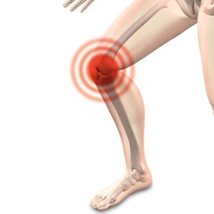
You may have swelling, aching and tenderness in the popliteal area of the knee.   The sac is located in the medial (inner) side of the popliteal fossa (diamond-shaped space behind the knee joint).


The Baker’s Cyst

The baker’s cyst is an idiopathic (cause unknown) form of bursitis where the cyst contents are not in the knee joint.

It can be found in children between ages 6-8 and 12-14 following rapid growth. Baker’s cysts in children are painless and normally disappear as the child matures. 

In contrast, a baker’s cyst found in adults is symptomatic. In adults it has intra-articular (within joint) problems such as degeneration of posterior horn of the medial meniscus (C-shaped cartilage between shin bone and thigh bone) or synovial membrane (connective tissue joint lining) irritation. 

Apparently, a baker’s cyst is due to swelling of the semimembranosus bursa.  The semimembranosus bursa is located between the semimembranosus (one of a group of muscles called the Hamstrings) and gastrocnemius (calf muscle) tendon.  Therefore, it is also called semimembranosus bursitis. Interestingly, poor flexibility of the semimembranosus and gastrocnemius muscles could contribute to development of a baker’s cyst. And this results in tendinitis.

Furthermore, degenerative joint disease or a torn meniscus are other possible contributing causes for a baker’s cyst.
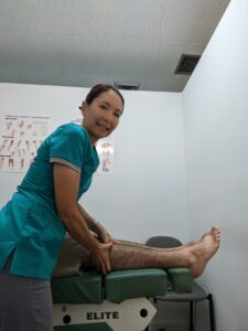
 

Treatment for a Baker’s Cyst at Meiri Chiropractic

First, Chiropractic care for the knee conditions (e.g. tendinitis) can resolve the cyst. 


Second, chiropractic manipulative therapy (adjustments) is rendered to the knee and associated spine and extremity (limb) joints.


Third, manipulation of muscle contractures with soft tissue techniques is utilized.  Some Techniques include gentle PNF/PIR (e.g. post isometric relaxation) and/or myofascial release techniques. 


Finally, therapeutic exercises for rehabilitation/ strengthening are prescribed. Especially, the lack of flexibility in the hamstrings and gastrocnemius are addressed.


Getting regular chiropractic treatment at a chiropractic center in West Palm Beach can help naturally relieve the pain and dysfunction of the knee. Chiropractic is a holistic and natural way to not only treat existing conditions, but to keep your body in its best working condition.

Contact Meiri Chiropractic today at 561-253-8984 to make an appointment or to learn more about  Knee Bursitis and Chiropractic Treatment: Baker’s Cyst.
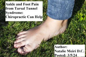


i


Do you have numbness and tingling across the bottom of the foot?  Furthermore, do you have burning pain in the ankle or foot?  It may be often at the sole or first toe. Your pain and paresthesias (numbness a tingling) probably came on insidiously (gradually).  And you do not have any associated low back or leg pain. You may also have noticed weakness in your foot. Lastly, your symptoms are worse by prolonged standing, walking, or running.  These are all symptoms of Tarsal Tunnel Syndrome.

In one study that evaluated patients with tarsal tunnel syndrome, 45 % reported numbness, 39% reported pain, and 16% reported both.  Read on to learn about Ankle and Foot Pain from Tarsal Tunnel Syndrome: Chiropractic Can Help.

Cause

The posterior tibial nerve may be stretched or compressed in the tarsal tunnel.  This is a tunnel formed by the flexor retinaculum (band of thickened deep fascia around tendons), calcaneus (large bone forming the heel), distal tibia (shin bone), and malleolus (inner and outer boney protuberances of ankle).

Contents of the tunnel include: the tendons of the posterior tibialis (muscle in posterior compartment of the lower leg), flexor digitorum longus (also located in posterior compartment muscles of lower leg), and flexor hallucis (muscle goes from calf to 1st toe) and the posterior tibial artery, vein, and nerve. 

Anatomy of the Tibial Nerve

Firstly, the tibial nerve is one of two main terminal branches of the sciatic nerve.  And the sciatic nerve is the largest nerve in the human body.

Secondly, the tibial nerve originates from the L4-S3 spinal nerve roots and provides motor and sensory innervation to most of the posterior (back part) leg and foot.

Thirdly, the tibial nerve descends from the popliteal fossa (space behind knee joint) into the posterior compartment of the leg. It passes deep to the fibular and tibial heads of soleus muscle (a calf muscle found between knee and heel). 

Fourthly, it runs vertically through the deep layer of the posterior (back) compartment of the leg, along with the posterior tibial blood vessels. The tibial nerve provides innervation to the surrounding muscles in this area.

Fifthly, the tibial nerve exits next at the ankle joint. It passes behind the medial malleolus (boney protuberance at inner ankle) to enter the sole of the foot.  Here it supplies innervation to most of the intrinsic (deep) muscles and skin.

Sixthly, past the tarsal tunnel, the tibial nerve gives off its terminal branches, the medial and lateral plantar nerves.  These nerves continue to supply motor and sensory innervation to the plantar foot.


Pronation of the Foot

Hyperpronation (excessive inward rotation of the foot during gait) is often the cause of tarsal tunnel syndrome.  This could be due to tightening of the flexor retinaculum or arch of the abductor hallucis muscle. This muscle is located on the inner border of the foot and bends the big toe and moves it out to the side. However, trauma and the subsequent swelling may also cause entrapment of the tibial nerve. 

There may also be a chronic tenosynovitis of the posterior tibial or flexor digitorum longus tendons.  This occurs near the malleolus (prominent bones at ankle) as areas that can compress the tibial nerve.  These fascial restrictions may be a primary cause. However, it is necessary to rule out ganglions, myositis ossificans, exercise compartment syndrome, and tumors.


Examination

Your doctor may order a nerve conduction study in a difficult unremitting case.  However, usually patients are diagnosed by the physical (orthopedic, neurological etc.) examination.


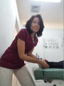
Dr Natalie Meiri adjusts a patient's ankle
Dr Natalie Meiri adjusts a patient’s ankle


Treatment at Meiri Chiropractic for Tarsal Tunnel Syndrome

First, soft tissue techniques will be rendered to the contracted fibrotic muscle and collagen at entrapment sites. Second, chiropractic adjustments (chiropractic manipulative therapy) to the foot, ankle and associated restricted joints along the kinetic chain is important.

Third, Therapeutic exercise will be given for stretching and strengthening.

Lastly, if trauma or swelling is involved, ice, compression, elevation, and physical therapy modalities is utilized to reduce swelling. Also, an orthotic and/or correction of shoes may be necessary if there is pronation of the foot.


Are your suffering from ankle or foot pain from Tarsal Tunnel Syndrome?

We offer excellent Chiropractic care for Tarsal Tunnel Syndrome. At Meiri Chiropractic, we spend the time necessary to examine, diagnose and treat every neuromusculoskeletal condition and various ailments you have. Indeed, Chiropractic is a holistic and natural way to not only treat foot and ankle pain, but to keep your body in its best working condition. We have been offering effective chiropractic care in West Palm Beach since 2006. Many of our patients reviews note our excellence. Call us today at 561-253-8984 to make an appointment or to find out more about Ankle and Foot Pain from Tarsal Tunnel Syndrome: Chiropractic Can Help.


Meiri Chiropractic

5601 Corporate Way, Ste#102

West Palm Beach, FL 33407


Many people dealing with Multiple Sclerosis (M.S.) feel as though they have very few options for managing their pain.   Chiropractic care maybe the answer!

In one study (1), manual chiropractic adjustive therapies appear to be responsible for the dramatic symptomatic relief provided for a patient diagnosed with M.S. Consequently, it was concluded “the relative risk-to-benefit ratio suggests that this approach may be appropriate as an alternative symptom management approach for MS”.


What is Multiple Sclerosis (M.S.)?

Typically, M.S. patients are less than 55. And it is more prevalent in women than men. MS appears in patients who reside in temperate climate zones and are of western European ethnicity. Relapses commonly occur in women 2 to 3 months after childbirth.

MS is characterized by episodic attacks that initially resolve, but eventually leave residual neurologic deficits. The initial episode often will resolve in days.  Next, the patient may remain symptom free for months or years. Eventually, however, symptoms recur.  Symptoms usually will involve a region and consist of numbness, tingling, weakness, diplopia (double vision), dizziness, or urinary sphincter dysfunction (urgency or hesitancy).

Cause

The cause of MS is unknown, but scientists think it may be an immune disorder (condition in which the body attacks itself by mistake).

There is an apparent genetic relationship due to the association of MS and HLA-DR2.  The human leukocyte antigen (HLA) is a family of genes found on chromosome 6 which provide the instructions to make a group of proteins known as the HLA complex. The HLA proteins are present on the surface of cells, and they help the immune system to distinguish foreign invaders such as viruses and bacteria from the body’s own cells and tissues. 

The primary role of the HLA complex is in the regulation of the immune system to defend against disease. Your individual HLA type is inherited from your parents. In MS, the immune system fails to distinguish between the body’s tissues and foreign proteins.  This results in your body attacking your own central nervous system. 


Demyelination

Firstly, MS attacks myelin as if it were foreign. And Myelin is an insulating layer, or sheath that forms around nerves, including those in the brain and spinal cord. Secondly, MS is a disease characterized by patchy demyelination with reactive gliosis.  Gliosis is a process leading to scars in the central nervous system that involves the production of a dense fibrous network of neuroglia (supporting cells) in areas of damage.  Thirdly, this occurs in the spinal cord, optic nerve (nerve for transmitting visual images), and white matter of the brain.  Lastly, Gliosis is a prominent feature of many diseases of the central nervous system, including multiple sclerosis and stroke.


Diagnosis and Findings on Imaging

No single test can diagnose MS. The medical history, neurologic exam and lab tests helps rule out other diseases and confirm MS. For example, Laboratory evaluation may reveal mild lymphocytosis (increased white blood cells) or increased protein count in the cerebrospinal fluid (CSF). Immunoglobulm G (type of antibody) are more often seen in the CSF.

Furthermore, Magnetic Resonance Imaging (MRI) will demonstrate multifocal areas of patchy demyelination in the brain or cervical (neck) spinal cord.  

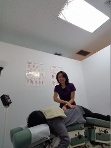
Dr. Natalie Meiri treating a patient

 
Management of Multiple Sclerosis

First, there is no cure for MS.

Second, natural Nutritional approaches may help, but there is not enough research.  Some studies suggest that an increase in polyunsaturated fatty acids and metabolic enzyme supplements help.  This nutritional supplementation may assist in providing an adequate lipid pool for oligodentrocytes (cell concerned with the production of myelin in the central nervous system). Also, antioxidants such as vitamins A, beta-carotene, E, C with bioflavinoids, and selenium may help with myelin membrane perioxidation (free radical reaction).  

Third, chiropractic is a popular CAM (complementary alternative medicine) therapy for symptom relief of M.S.  In one study, conservative chiropractic manual adjustive therapies were provided to address spinal segmental dysfunction.  And the patient reported complete absence of symptoms following the first treatment intervention.

Are you seeking a natural treatment to help relieve Multiple Sclerosis (MS) symptoms?  Your chiropractor in West Palm Beach can help you manage your symptoms and improve your overall quality of life. Call 561-253-8984 today to make an appointment or to find out more about Chiropractic Care in West Palm Beach: Multiple Sclerosis Pain Relief.


If you have knee pain from a collateral ligament injury, Chiropractic can provide relief! This post is about Knee Pain and Chiropractic Treatment: Collateral Ligament Injury.


Anatomy of the knee: Femur (thigh bone), Tibia (shin bone), Fibula (thin, outer long bone of the leg), and the Patella (kneecap)

There are three “articulations” (joint or juncture between bones or cartilages) at the knee: the tibiofemoral, patellofemoral, and tibiofibular articulations. However, only the tibiofemoral and patellofemoral articulations participate in knee joint activity. The tibiofibular articulation/joint does not actually contribute to the actions of the knee. Instead, it is part of the ankle joint complex moving the ankle in all the various ranges of motion. However, dysfunctional processes in the proximal tibiofibular articulation/joint can affect other knee functions and can be a source of knee pain.

So the tibiofemoral and tibiofibular are synovial articulations.  These are freely movable joints. And the bony surfaces are coated with hyaline cartilage.  It is unified by a fibrous articular capsule. However, the third articulation, patellofemoral, is a functional joint (non-synovial joint).   

 More Anatomy of the Knee

First, the knee is supported by the joint capsule, its cartilage “thickenings”, and the collateral-ligamentous system.  And the collateral ligaments (band of tissue that connects a bone to another bone) are located on either side of your knee outside your knee joint.

Second, internally the knee is stabilized and the control of rotation is provided by the meniscocruciate system. There are two C-shaped cartilage like structures, each one called a meniscus. The menisci aid in shock absorption and help govern rotational movement at the knee.

Third, the cruciates, capsule, and collateral ligaments connect the femur to the tibia. The cruciates are a pair of ligaments that cross each other in the knee, connecting the tibia and the femur. The Anterior Cruciate Ligament (ACL) is smaller (size of the little finger) than the Posterior Cruciate Ligament.  It’s usually more readily injured than the Posterior Cruciate Ligament (PCL) (size of the thumb). The cruciates allow tension to develop through most ranges of motion. 


Cause of Pain in the Knee due to Collateral Ligament Injuries

Firstly, the knee is affected by a variety of conditions.  However, a traumatic event is by far the most common. The MCL (medial collateral ligament) is the most commonly injured knee ligament, and LCL (lateral collateral ligament) injuries are usually associated with more severe knee injuries.  So injuries to the collateral ligaments usually require some type of traumatic force.

Secondly, in external rotation of the knee, your toes go outward and your heel goes inward.  A frequent trauma occurs with a twisting external rotation movement of the knee while flexed (bent).  Furthermore, a valgus/abduction (hit from outside of knee) blow to the knee is another common mechanism for MCL injuries. In contrast, a LCL injury may occur with a varus/adduction (hit from inside of knee) blow to the knee. 

Thirdly, the knee joint is innervated (supplied) by spinal nerve segments L3–S1.  Therefore, in cases of pain of nontraumatic onset, problems elsewhere in (e.g. segments L3–S1) must be ruled out. Lastly, the lumbar spine, hip, and foot are possible sources of referred pain to the knee as well. 

Fourthly, chronic strain through valgus-loading of the knee (e.g., pronation) can cause nontraumatic injury. Ultimately, this repetitive stress injury can lead to a collateral ligament sprain.

Finally, your doctor should take all the above into account when examining, diagnosing and then treating your knee.  Of course, you will require imaging such as an x-ray or/and M.R.I. to properly diagnose your knee condition.


Collateral Ligament Injury Grades

With first-degree tears, you will frequently not have any history of trauma.  You may report more mild to moderate pain at the knee. And overuse activity may have brought on this knee sprain.

When the tear is second or third degree, there is usually a traumatic incident as described above. You will have sharp knee pain with associated swelling at the time of injury.


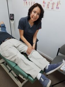
Dr. Natalie Meiri adjusts a patient's knee
Dr. Natalie Meiri adjusts a patient’s knee


Knee Pain and Chiropractic Treatment: Collateral Ligament Injury

First, chiropractic manipulative therapy is rendered to the knee and associated spine and extremity (limb) joints.


Second, manipulation of muscle contractures with soft tissue techniques is utilized.  Some Techniques include gentle PNF/PIR (e.g. post isometric relaxation) and/or myofascial release techniques. 


Finally, Therapeutic exercises for rehabilitation/ strengthening are prescribed. 


Getting regular chiropractic treatment at a chiropractic center in West Palm Beach can help naturally relieve the pain and dysfunction of the knee. Chiropractic is a holistic and natural way to not only treat existing conditions, but to keep your body in its best working condition.

Contact Meiri Chiropractic today at 561-253-8984  to make an appointment on Knee Pain and Chiropractic Treatment:  Collateral Ligament Injury or/and other ailments.


If you have knee pain from an anterior cruciate ligament injury, Chiropractic can provide relief! This post is about Knee Pain and Chiropractic Treatment: A.C.L. Injury.


Acute Phase of  the Anterior Cruciate Ligament (ACL) Injury

If you are in the acute phase, you may have sudden knee pain after a “contact injury”.  Your knee may  get hit very hard on the side. In addition, it may get hit while in a hyperextension (knee bent backwards) position.  Furthermore, you may have heard a “pop” during the injury.  Next, your joint will swell and you won’t be able to weight bear. 


Chronic Phase of the Anterior Cruciate Ligament (ACL) Injury

If you are in the chronic phase, you may have had the above happen in the past.  Of course, it may have resolved gradually after some swelling and pain.  Finally, you may feel more of an instability than pain in your knee.  Moreover, you may not even remember what happened. So this would be a noncontact/ nontraumatic ACL injury.  And this often occurs when an athlete slows down, pivots while running, and lands from a jump. Typically, these movements are common in sports, especially in soccer, basketball, and football.


Anatomy: Femur (thigh bone), Tibia (shin bone), Fibula (thin, outer long bone of the leg), and the Patella (kneecap)

There are three “articulations” (joint or juncture between bones or cartilages) at the knee: the tibiofemoral, patellofemoral, and tibiofibular articulations. However, only the tibiofemoral and patellofemoral articulations participate in knee joint activity. The tibiofibular articulation/joint does not actually contribute to the actions of the knee. Instead, it is part of the ankle joint complex moving the ankle in all the various ranges of motion. However, dysfunctional processes in the proximal tibiofibular articulation/joint can affect other knee functions and can be a source of knee pain.

So the tibiofemoral and tibiofibular are synovial articulations.  These are freely movable joints. And the bony surfaces are coated with hyaline cartilage.  It is unified by a fibrous articular capsule. However, the third articulation, patellofemoral, is a functional joint (non-synovial joint).   


More Anatomy of the Knee: About the ACL

First, the knee is supported by the joint capsule, its cartilage “thickenings”, and the collateral-ligamentous system.  And the collateral ligaments (band of tissue that connects a bone to another bone) are located on either side of your knee outside your knee joint.

Second, internally the knee is stabilized and the control of rotation is provided by the meniscocruciate system. There are two C-shaped cartilage like structures, each one called a meniscus. The menisci aid in shock absorption and help govern rotational movement at the knee.

Third, the cruciates, capsule, and collateral ligaments connect the femur to the tibia. The cruciates are a pair of ligaments that cross each other in the knee, connecting the tibia and the femur. The Anterior Cruciate Ligament (ACL) is smaller (size of the little finger) than the Posterior Cruciate Ligament.  It’s usually more readily injured than the Posterior Cruciate Ligament (PCL) (size of the thumb). The cruciates allow tension to develop through most ranges of motion. 


Complications and Causes of Knee Pain from the ACL Injury

An anterior cruciate ligament injury is the over-stretching or tearing of the anterior cruciate ligament (ACL) in the knee.

As mentioned previously, ACL damage can be from a noncontact injury or a sudden traumatic contact event.   Usually, excessive contraction of the quadriceps muscle with insufficient hamstrings co-contraction occurs with sudden stopping or cutting manuevers.  Also, contact injury with a rotary component is more likely to damage the ACL and other structures such as the menisci or MCL (medial collateral ligament).

When the ACL tears, it usually occurs in the mid substance of the ligament. And because of the closeness of the vasculature, blood vessel rupture often occurs.  Lastly, this causes quickly developing joint swelling that is both tense and painful. 


ACL injury Grade 1-3

Sprain is grade 1– microscopic tears that stretch the ligament.

Partial ACL tear is grade 2– stretched and partially torn.

Complete ACL tear or an avulsion is grade 3- the Tibial spine avulsion ACL injury occurs in teens. The ACL hasn’t torn, but the attachment of the ligament to the tibia has pulled off partially or entirely.  With a complete tear you may need surgery.  However, you should always try conservative therapy/treatments such as chiropractic first.


Dr. Natalie Meiri adjusts a patient's knee
Dr. Natalie Meiri adjusts a patient’s knee


Evaluation, Tests and Management

After examining your knee, your doctor may order imaging such as an x-ray or/and M.R.I. Grade 1 ACL injuries may not require bracing.  However, second to third grade tears (without involvement of the meniscus) may require a month of restricted bracing.


Knee Pain and Chiropractic Treatment: A.C.L. Injury Treatment at Meiri Chiropractic

First, the knee and associated spine and extremity (limb) joints are adjusted.


Second, manipulation of muscle contractures with soft tissue techniques is utilized.  Some Techniques include gentle PNF/PIR (e.g. post isometric relaxation) and/or myofascial release techniques. 


Finally, Therapeutic exercises for rehabilitation/ strengthening are prescribed. 


Getting regular chiropractic treatment at a chiropractic center in West Palm Beach can help naturally relieve the pain and dysfunction of the knee. Chiropractic is a holistic and natural way to not only treat existing conditions, but to keep your body in its best working condition.

Contact Meiri Chiropractic today at 561-253-8984  to make an appointment on Knee Pain and Chiropractic Treatment:  A.C.L. Injury or/and other ailments.


If you have knee pain from osteoarthritis (wear and tear arthritis), Chiropractic can provide relief! This post is about Knee Pain and Chiropractic Treatment: Knee Arthritis

You may have stiffness and knee pain. Your knee pain may be worse with prolonged sitting and walking.  Moreover, you may have a history of trauma or surgery to your knee.  Additionally, you may have recurrent bouts of swelling and “bowlegged” appearance to the knee.


The Femur (thigh bone), Tibia (shin bone), Fibula (thin, outer long bone of the leg), and the Patella (kneecap)

There are three “articulations” (joint or juncture between bones or cartilages) at the knee: the tibiofemoral, patellofemoral, and tibiofibular articulations. However, only the tibiofemoral and patellofemoral articulations participate in knee joint activity. The tibiofibular articulation/joint does not actually contribute to the actions of the knee. Instead, it is part of the ankle joint complex moving the ankle in all the various ranges of motion. However, dysfunctional processes in the proximal tibiofibular articulation/joint can affect other knee functions and can be a source of knee pain.

So the tibiofemoral and tibiofibular are synovial articulations.  These are freely movable joints. And the bony surfaces are coated with hyaline cartilage.  It is unified by a fibrous articular capsule. However, the third articulation, patellofemoral, is a functional joint (non-synovial joint).  Nevertheless, many of the same problems such as arthritis arise here.


More Anatomy of the Knee

First, the knee is supported by the joint capsule, its cartilage “thickenings”, and the collateral-ligamentous system.  And the collateral ligaments (band of tissue that connects a bone to another bone) are located on either side of your knee outside your knee joint.

Second, internally the knee is stabilized and the control of rotation is provided by the meniscocruciate system. There are two C-shaped cartilage like structures, each one called a meniscus. The menisci aid in shock absorption and help govern rotational movement at the knee.

Third, the cruciates, capsule, and collateral ligaments connect the femur to the tibia. The cruciates are a pair of ligaments that cross each other in the knee, connecting the tibia and the femur. The Anterior Cruciate Ligament (ACL) is smaller (size of the little finger) than the Posterior Cruciate Ligament.  It’s usually more readily injured than the Posterior Cruciate Ligament (PCL) (size of the thumb). The cruciates allow tension to develop through most ranges of motion.


Causes of Knee Pain from Osteoarthritis

Degeneration of articular (joint) cartilage is often secondary to meniscal tearing or degeneration. Unfortunately, this occurs with age. Additionally, single trauma events or a past knee surgery (including ACL or meniscal surgeries) predispose you to early degeneration.


What is Osteoarthritis (Degenerative Joint Disease)?

Degenerative arthritis or degenerative joint disease is also referred to as osteoarthritis.  It is a “wear and tear” arthritis. This serious, painful condition can affect any joint of the body (e.g. hands, knees, hips, shoulders, back and neck). It is by far the most common form of arthritis, affecting more than 32.5 million adults in the United States, according to the Centers for Disease Control and Prevention.

Firstly, osteoarthritis is a disease of the entire joint, including bone, cartilage, ligaments, menisci (e.g. knee meniscus) and the tissues lining the joint (the synovium). There can also be changes in periarticular (around the joint) muscles, nerves, bursa, and local fat pads that may contribute to the symptoms of osteoarthritis.

Secondly, it degrades cartilage, changes bone shape and this causes inflammation. 

Thirdly, the result is pain, stiffness and loss of mobility.

Lastly, pain and other symptoms of osteoarthritis may lead you to feel tired, have problems sleeping, and possibly feeling depressed.


Options for managing Osteoarthritis along with chiropractic care:

-Exercise is an effective management approach to OA of the knee with significant reductions in pain and improvements in function. 

– Taping and bracing may also be effective management approaches. A recent blinded, randomized controlled trial was conducted to evaluate the effectiveness of knee tape. Those who were given therapeutic taping had significant improvement in pain.These effects were maintained after three weeks. Another study designed to evaluate the effectiveness of an elastic knee brace produced results that indicate small, short-term beneficial effects with acute exacerbations.

-Use of glucosamine and chondroitin supplementation appears to have both an effect on symptoms and structural changes in the knee. The results from a recent meta-analysis indicate that both glucosamine and chondroitin demonstrated improvements in joint space narrowing.


Dr. Natalie Meiri adjusts a patient's knee
Dr. Natalie Meiri adjusts a patient’s knee


Research on How Chiropractic Adjustments Can Help With Arthritis

A 2017 review in the Journal of the American Medical Association, found that spinal manipulation reduces lower back pain. And a 2013 study published in Osteoarthritis and Cartilage found that patient education combined with 12 chiropractic treatments (twice a week for six weeks) were more effective for hip Osteoarthritis than a daily stretching program or patient education alone. 

Chiropractic Care of Degenerative Arthritis of the Spine and Extremities at Meiri Chiropractic:

Typically, our program includes Chiropractic Manipulative Therapy to the spine and extremity (arms and legs) joints for 2-3 months. Then “maintenance of normal joint motion and function of the spine and extremities may be facilitated by adjusting/manipulation” (1).  

Also, manipulation of muscle contractures with soft tissue techniques may be accomplished with gentle PNF/PIR (e.g. post isometric relaxation) and/or myofascial release techniques. Next, therapeutic exercises for strengthening of the joint will help for active care at home.   Finally, recommendations on an anti-inflammatory diet will be of benefit for you.

Getting regular chiropractic treatment at a West Palm Beach chiropractic center can help naturally relieve the pain and dysfunction of degenerative arthritis of the knee. Chiropractic is a holistic and natural way to not only treat existing conditions, but to keep your body in its best working condition.

Contact Meiri Chiropractic today at 561-253-8984  to make an appointment on Knee Pain and Chiropractic Treatment: Knee Arthritis or/and other ailments.


References:

(1) Thomas Bergman, David Peterson, Chiropractic Technique Principles and Procedures, 3rd edition, 2011


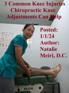
When thinking of chiropractic, people always think it’s only for the spine.  Moreover, the question people ask often is, “can a chiropractor adjust knees?”  The answer is, yes. In fact, it is an effective natural treatment option for those suffering from many conditions leading to knee pain.  Chiropractic adjustments can help with knee pain by improving joint function, thereby providing relief.

Is knee pain affecting your life and causing discomfort and limiting mobility? In this post we will explore 3 Common Knee Injuries Chiropractic Knee Adjustments Can Help.


1.  Knee Meniscus Tears

The knee meniscus is the rubbery knee cartilage that cushions the tibia (shinbone) from the femur (thighbone).

You may have knee pain usually following a rotational injury to the knee. Next, there may be swelling in your knee that developed over a number of hours. And since the injury, you may have episodes of knee locking wherein you experience significant pain.  Also, you may not be able to move the leg for a few seconds until the injured knee is forcefully extended (straightened). Furthermore, you may complain of the knee “giving way”.


Imaging and Evaluation

There are examination (e.g. orthopedic/neurologic/chiropractic ) tests that have a positive finding for a meniscal tear. Of course, Meniscal Tears are seen on a magnetic resonance imaging (MRI).  However, clinical correlation (results of the test should be interpreted in conjunction with the patient’s clinical symptoms, medical history, and other relevant diagnostic information) with MRI findings are important.  This is because there may be other pathologies (cause of the disease) causing pain as well.


Cause of Meniscus Tears

Firstly, most meniscus injuries are due to combined compression with rotation at the knee. Secondly, there are many types of tears. They are generally divided into horizontal and vertical tears. Thirdly, because of the minor blood supply, meniscus tears do not bleed much into the joint. However, the free edge of the tear may cause irritation along with the synovial (thin membrane of joint) fluid production. Fourthly, the meniscus is often involved when the anterior cruciate ligament (ACL) is torn.


2.  Popliteus Tendinitis

First, the popliteus muscle lies behind the knee joint. It forms the base of the popliteal fossa (diamond-shaped space behind the knee joint). Second, it inserts at the lateral femur, lateral meniscus and inserts onto the proximal (nearer to the center of the body) tibia (shin bone). Third, the popliteus muscle does lateral rotation of the femur relative to the tibia. And this ‘unlocks’ the knee joint so that the knee can flex (bend). Fourth, Popliteus is a major stabilizing muscle of the knee.  Fifth, tendinitis is a condition in which the tissue connecting muscle to bone becomes inflamed.

You will have pain in the lateral (side away from body) knee following downhill running or walking. Your foot on the injured side may have hyperpronation problems. 

 
3.   ProximaI TibiaI-Fibular Chiropractic Subluxation (chiropractic intersegmental dysfunction/misalignment)

The tibial-fibular joint is formed by the tibia and the fibula (outer of the two bones between the knee and the ankle). The proximal tibial-fibular articulation is a synovial joint (freely movable joint). Synovial joints are the main functional joints of the body.  Synovial joint cavities contain synovial fluid secreted by the synovial membrane (synovium), which lines the articular capsule.

Movement at this joint is influenced by movement at the ankle.  Sudden forced movements (in particular dorsiflexion of ankle) may force the fibula into a fixed position. With hamstring curls, the biceps femoris (one of heads of hamstrings) may draw the fibular head posteriorly (back), leading to a chiropractic subluxation/misalignement.  If left untreated, it may lead to entrapment of the peroneal nerve by the fibula.  So compression or entrapment of the nerve may occur with the result of radiating pain down the outside lower leg.


3 Common Knee Injuries Chiropractic Knee Adjustments Can Help

The chiropractic approach to the treatment of knee injuries involves a thorough understanding of the anatomy, biomechanics, motor patterns, and kinetic chains of the lower extremity (limb), including the pelvis. Your kinetic chain is a combination of several successively arranged joints. These joints constitute a complex unit, as links in a chain.

By identifying the underlying problem or dysfunction, your chiropractor can implement a multilevel treatment protocol (detailed plan) that involves chiropractic manipulative therapy.  A chiropractic realignment will restore faulty biomechanics. Lastly, your chiropractor can prescribe exercises to strengthen weakened muscle groups and motor pattern re-education.

At Meiri Chiropractic we provide chiropractic adjustments/ manipulation for your knee and any associated regions (e.g. foot, ankle, hip, pelvic or lumbar (low back) indicated.  Additionally, soft tissue therapy (myofascial release, pressure point and various relaxation procedures) and modalities (cold/heat therapy and electric muscle stimulation) are rendered as necessary.

Do you know someone who needs Chiropractic adjustments for a knee injury? Contact Meiri Chiropractic serving West Palm Beach, Jupiter, North Palm Beach, and Palm Beach Gardens today at 561-253-8984 for this or some other ailment.


    Video Vlog on same post


A common source of joint fixation/chiropractic subluxation (hypomobility) and dysfunction is periarticular (surrounding joint) soft tissue injury with its resultant fibrosis (thickening or scarring of the tissue) and loss of elasticity and strength. So soft tissue injury and fibrosis may result from trauma to muscular, tendinous, myofascial, or ligamentous tissue. And you may have had acute trauma or repetitive injury to the area to cause it. 


Chiropractic Adjustments Restore Motion


First, an inflammatory response is triggered resulting in an extracellular (taking place outside of cell) accumulation of exudates (fluid that leaks out of blood vessels) and blood. Platelets (tiny blood cells that help your body form clots to stop bleeding) then release thrombin (molecule which helps in clotting process) converting fibrinogen into fibrin, which organizes into collagenous scar tissue. This results in a variety of soft tissue and articular (joint) adhesions.


Second, the above response can often be excessive especially in traumatic neuromusculoskeletal injuries. Therefore, therapies for minimizing inflammatory exudates are helpful.  This will in turn reduce pain and muscle spasm. Ultimately, this leads to early pain-free mobilization and flexible repair.


Third, early care and mobilization provide the best opportunity for optimal healing.

In contrast, bed rest and prolonged inactivity increases the chances of long-term disability and eventually lost time from work.


Fourth, the exudates that arise due to injury and inflammation gets the next step ready of connective tissue repair. Moreover, the matrix for the development of granulation tissue (new connective tissue) and scar formation is set. And the formation of granulation tissue is mostly carried out by the rapid increase of fibroblasts (cells that contribute to the formation of connective tissue).  Next, fibroblasts synthesize and deposit collagen tissue. At first, the collagen is poorly organized.  And additional collagen cross-linkages to reorganize along planes of stress which improves the tensile (stretchability) strength of the injured area sets forth.


Fifth, this repair and remodeling process may take months and may result in less than optimal healing and extensibility (stretchability) of the involved tissue.  Of course, immobilization slows recovery. This results in a loss of strength and flexibility and possible intra-articular (within the joint) fatty adhesions.  Immobilization also leads to dehydration, causing proteoglycans (substance which provides hydration and swelling pressure to the tissue enabling it to withstand compressional forces) to get closer and stick together. 


In conclusion, if injury or immobilization leads to decreased flexibility, therapies such as chiropractic adjustments are directed toward the restoration of motion.


Dr. Natalie Meiri adjusts a patient


Myofascial Cycle of Pain


Myofascial refers to the muscle or of the fascia (layer of connective tissue that supports tissues and organs) surrounding the muscleSo myofascial conditions are triggered by many reasons.  This can become self-perpetuating sources of pain, muscle spasm, and joint dysfunction.

Additionally, painful conditions which cause chronic muscle hypotoncity (deficient tone) are additional origins of restricted joint motion. In other words, muscle contraction, once initiated, may become a self-perpetuating source of pain and muscle hypotoncity.  Protective splinting in the joint’s intrinsic muscles may further this process by stopping passive joint movement and the pain-inhibiting qualities of certain joint receptors.

If contractions continue, over time they may develop into muscle contractures.  This is a result of adaptational shortening and loss of elasticity from disuse or underuse. Problems capable of producing muscle contraction are: trauma, structural inadequacies, visceral (organ) disease, emotional distress, and exposure to cold.


Myofascial Cycle Summary


  1. Joint Dysfunction leads to muscle strain.
  2. Muscle strain is caused by: Repetitive use, Emotional tension, Chronic postural stress, Exposure to cold, Visceral disease, Physical trauma, structural inadequacies, uncoordinated movements
  3. Vasoconstriction (narrowing of blood vessels by small muscles in their walls) and ischemia (inadequate blood supply) is caused by the pain of muscle strain. So pain causes muscle splinting. This in turn leads to retained metabolites and edema (inflammation).
  4. Sustained contraction happens due to vasoconstriction/ischemia and along the way myofascial syndromes and joint dysfunction occur.
  5. Fibrous reaction occurs finally and leads to soft tissue contractures.
  6. And Soft tissue contractures lead to Persistent joint and somatic (skin, muscles, joints, and bones) dysfunction as the end result.

 
Some Causes of Chiropractic Subluxations: Stopping the Myofascial Cycle


Behaviors or bad habits can cause subluxations.  For example the following may contribute: continuously carrying a purse or bag on the same side of your body, sleeping on your stomach, sleeping with a pillow that is too big or too flat for you, crossing your legs or looking down at a computer monitor for long periods of time, sitting for long hours, and improper lifting. Also, de-stressing and decreasing your exposure to cold (e.g. excessive air conditioning) may help. 

Meiri Chiropractic utilizes the best joint manipulation and adjustments to the spine and peripheral joints. We provide soft tissue procedures, hot and cold modalities, stretching/ rehabilitation exercises, and homeopathic consultations as well.

At Meiri Chiropractic in West Palm Beach we spend the time necessary to examine, diagnose and treat every neuromusculoskeletal condition and various ailments you have. Through regular chiropractic visits, you can get pain relief and improve function. Chiropractic is a holistic and natural way to treat existing conditions.  And your body will be in its best working condition with regular chiropractic care.  Meiri Chiropractic has been offering effective chiropractic care in Palm Beach county since 2006.  Many of our patient reviews note our excellence.  Call us today at 561-253-8984 in West Palm Beach to make an appointment.


References: Thomas Bergman, David Peterson, (3rd edition) Chiropractic Technique: Principles and Procedure


From Dr. Natalie Meiri’s Clinical Pearl Stories: Chiropractic Care for Mid-Back and Right Hand Pain Over the Holidays


This was a 45 year old patient who was getting ready for the Christmas holidays. To be HIPAA compliant, I will call her Kathlyn instead of her real name.  Kathlyn was at a holiday party when a “7 foot tall enormous man” gave her a “bear hug”.  She stated “although I was happy to see my friend, the hug injured my spine”.   To make matters worse, she was getting her Christmas gifts ready when she somehow injured her right hand. She presented to my office the next day with mid back and right hand pain.

Kathlyn hadn’t gone to urgent care or her primary care, just took some NSAIDS (Non-steroidal anti-inflammatory drugs) without much relief.   On the intake form she reported pain in her midback and right hand.


Kathlyn’s Examination and Treatment

Upon examination, there were positive tests for her thoracic spine (midback) as well as the right hand/fingers.  After that, I explained her diagnosis (strain of thoracic spine and sprain of right hand), prognosis and treatment plan.

Firstly, Kathlyn’s chiropractic treatment for thoracic strain and right hand sprain consisted of chiropractic manipulation/ adjustments of the thoracic spine, right hand, and associated regions.  Associated regions that were adjusted were her cervical spine (neck), lumbar spine (low back) and right wrist which were misaligned (subluxations or intersegmental dysfunctions). Secondly, soft tissue therapy (myofascial release, pressure point and various post isometric relaxation procedures) and modalities (cold/heat therapy and electric muscle stimulation) were administered as needed. Thirdly, she was given therapeutic exercises to continue her treatment at home. Fifthly, Kathlyn was given a brace to wear during strenuous activities for her right hand.


What is a strain?

A strain is an overstretching and tearing of musculotendinous tissue. Tendons are connective tissue that attach a muscle to other body parts, usually bones.


What is a sprain?

A sprain involves a ligament.  It is a short band of tough, flexible fibrous connective tissue.  Furthermore, it connects two bones together in proper articulation at the joints.

A Joint injury in which some of the fibers of a supporting ligament are torn or ruptured is a sprain.  However, the continuity of the ligament remains intact in a sprain.  Grade 1 (mild) sprains cause slight stretching and tiny tears in the ligament. Kathlyn had a grade 1 sprain of her right hand. Grade 2 (moderate) sprains refer to partially torn ligaments with pain and swelling.


Kathlyn’s Outcome

The outcome for Kathlyn was excellent! She felt a little better immediately from the first visit.  Moreover, after only a few visits she enjoyed the rest of the holiday season pain-free.


Chiropractic Care for Mid-Back and Right Hand Pain Over the Holidays

Many people “throw their back out” every year around the holidays. This could be due to stress or “holiday bear hugs” as in the case of Kathlyn. Chiropractic is powerful and great at com-batting the negative effects of stress on the body. Adjustments restore joint mobility and function, resolves joint inflammation and reduces the patient’s pain.

Do you know someone who is in need of chiropractic care this holiday season? Contact Dr. Natalie Meiri of Meiri Chiropractic today at 561-253-8984 to learn more about Chiropractic Care for Mid-Back and Right Hand Pain Over the Holidays or to schedule an appointment.


Your feet and ankles are important for the balance of your whole body. Not only do your feet and ankles provide support, they absorb shock.  The stability of your feet and ankles are important for walking, running and jumping. And your feet and ankles have to provide mobility while adapting to different terrain.

Shock absorption is distributed through complex movements through your kinetic chain.  So these forces from various movements are distributed through the lower extremity from your feet/ankle to the knee, hip, and then the pelvis. Therefore, dysfunction at your ankles or feet may have consequences throughout your entire body. Furthermore, your foot problems may be local or referred.  In other words, the need for lower-extremity compensation may result in pain somewhere else, including low back pain.


Problems in the Feet

Most foot complaints are due to lack of proper support or inappropriate footwear. Ill-fitting or irritating shoes may result in corns or calluses, sesamoiditis (inflammation of the sesamoid bones in the ball of the foot and the tendons they are embedded in) or aggravation of hallux valgus (bunion) at the first toe, or fat pad syndrome at the heel . Moreover, tight-fitting shoes may also cause compression of metatarsals (5 forefoot bones).

Also, if the foot is too mobile (i.e., hyperpronated), there may be a lack of support.  This may cause plantar fasciitis and strain of the tibialis posterior muscle (deep calf muscle). Indeed, proper shoes are important.  And if you have any forefoot (front of foot) or hind foot (back of foot) abnormalities (i.e. varus or valgus) you are more likely to strain your feet.  

Complicating Factors

Firstly, as the furthest part of your body, your foot is commonly affected by vascular disorders. Arterial occlusive (inadequate blood flow due to narrowing of the arteries) disorders block blood flow. Valvular insufficiency in veins along with gravity may lead to vascular stasis (veins can’t send blood back to heart).

Secondly, neurologic dysfunction associated with metabolic neuropathies can occur.  For instance, as seen with diabetic neuropathy.

Thirdly, being bare footed may lead to trauma. In contrast, the supported foot is susceptible to the pressure effects and biomechanical alterations of footwear. Especially if you are diabetic this can lead to unnoticed and untreated problems due to sensory deficits (i.e. touch, temperature, pain and other sensations).

Common Conditions of the Foot and Ankle

Some common conditions involving the foot and ankle that are biomechanical in nature include the following:

  1. First toe-hallux valgus/rigidus (stiff first toe), turf toe (sprain to the main joint of the big toe), and sesamoiditis
  2. Metatarsalgia (pain and inflammation in the ball of your foot)
  3. Peroneal tendinitis (inflammation in the tendons that run along your outer ankle/foot)
  4. Plantar fasciitis (inflammation of fascia tissue on the bottom of your feet)
  5. Achilles tendon/heel-tendinitis, bursitis (painful inflammation of small, fluid-filled sacs that cushion the bones, tendons and muscles near your joints) and fat pad syndrome
  6. Neuropathy

In addition to the above, ankle inversion and eversion sprains are very common. The most common are plantar flexion/inversion (foot turned inward) sprains. Also, chronic ankle pain and/or instability is not uncommon following repeated ankle sprains.

It is important not only to treat the acute injury, but also to prevent future occurrences. This often entails evaluation of the activity or sport coupled with a post injury rehabilitation program.


Chiropractic for Ankle and Foot Pain and Conditions at Meiri Chiropractic in West Palm Beach

Are your suffering from ankle or foot pain? We offer excellent Chiropractic adjustments/manipulation to the ankle, foot and associated areas (i.e. knee, hip, pelvis). Soft tissue techniques along with the use of modalities (i.e. heat therapy, electric muscle stimulation) and rehabilitation/home exercises are utilized along with the adjustments to get you pain-free.

At Meiri Chiropractic, we spend the time necessary to examine, diagnose and treat every neuromusculoskeletal condition and various ailments you have. Chiropractic is a holistic and natural way to not only treat foot and ankle pain, but to keep your body in its best working condition. We have been offering effective chiropractic care in West Palm Beach since 2006. Many of our patients reviews note our excellence. Call us today at 561-253-8984 to make an appointment or to find out more about Chiropractic for Ankle and Foot Pain and Conditions.