From Dr. Natalie Meiri’s Clinical Pearl Stories: Chiropractic Care of Facial Numbness and Neck Pain After a Car Accident

Author: Natalie Meiri, D.C.        Posted: 7/30/22


This was a 50 year old male patient with a chief complaint of acute left facial numbness and cervical (neck) and cheek pain following a rear-end car accident a few weeks ago. To be HIPAA compliant, I will call him Joe instead of his real name. When he was rear ended during the collision, he didn’t hit his head or loose consciousness.  However, his neck was “whipped” back and forth violently.


Joe never had facial numbness or cheek and neck pain prior to his car accident. Now Joe had trouble sleeping and couldn’t exercise or play with his kids due to his pain. Furthermore, he had difficulty working and had to take time off. 

 Joe’s Examination and Imaging

 Upon examination he had painful spasmed muscles and tenderness all around the cranium (head) and cervical (neck) spine. His sensory examination showed a pattern of numbness in his face due to a left sided trigeminal neuropathy. Joe had positive tests to indicate he had not only sprained and strained his neck, but also strained (irritate) his trigeminal nerve. And his cervical (neck) x-rays taken in my office showed loss of cervical lordosis (staightening) consistent with cervical myospasm and a sprain.  If he wasn’t responding to care in a month, I would order a M.R. I. (magnetic resonance image).

The Trigeminal Nerve

The trigeminal nerve is the part of the nervous system responsible for sending pain, touch and temperature sensations from your face to your brain.  You have two trigeminal nerves, one on each side of your head. The nerve has three divisions: the ophthalmic, maxillary, and mandibular nerves.

It is the 5th of 12 cranial nerves. The trigeminal nerve primarily helps you feel (sensory), although the mandibular branch of the trigeminal nerve has both sensory and motor functions. It helps with biting, chewing, swallowing, and facial and scalp sensations.

The trigeminocervical nucleus is a region of the upper cervical (neck) spinal cord where sensory nerve fibers in the descending tract (nerve pathways from face to brain) of the trigeminal nerve called the trigeminal nucleus caudalis are believed to interact with sensory fibers from the upper cervical nerve roots. This functional convergence of upper cervical and trigeminal sensory pathways allows the bidirectional referral of painful sensations between the neck and trigeminal sensory receptive fields of the face and head.


Dr. Natalie Meiri adjusts a patient


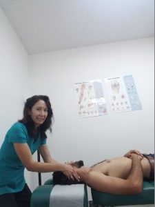
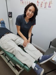
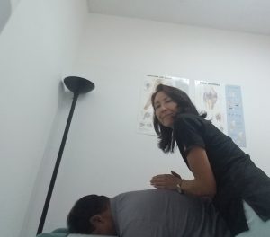
Joe’s Treatment at Meiri Chiropractic

It is common to have numbness and/or pain in your arm and neck pain after a car accident.  Similarly, you can have numbness in your face and have neck and left cheek pain after a car accident as Joe did. 

In Joe’s case, the strain (irritation) on the trigeminal nerve likely occurred at the upper cervical spine, at the nerve’s caudalis, and at the brainstem (bottom, stalk like portion of your brain connecting your brain to your spinal cord). Also, the temporal portion of his cranium (side of the head behind the eye between the forehead and the ear) needed manipulation due to the irritation of the maxillary branch of the trigeminal nerve.  Furthermore, the irritation of the maxillary branch of the trigeminal nerve which is responsible for sensations in the middle part of your face (e.g.cheeks, nose lower eyelids, upper jaw) must have caused his cheek pain.

Chiropractic manipulative treatment techniques applied at the level of the cervical spine, suboccipital (back of the skull) region, and cranial (head) region alleviated Joe’s facial symptoms. This treated the left-sided neuropathy of the trigeminal nerve and the neck sprain/strain.

Our other therapies, such as soft tissue techniques, electric muscle stimulation, ice/ heat therapies and homeopathic consultations provided Joe additional relief. It took about 4 months of care, but the facial numbness and neck and cheek pain subsided. Joe was able to heal without drugs or surgery!


We can help you with Chiropractic Care of Facial Numbness and Neck Pain After a Car Accident

At Meiri Chiropractic we spend the time necessary to examine, diagnose and treat every neuromusculoskeletal condition and various ailments you have.  Chiropractic is a holistic and natural way to not only treat existing conditions, but to keep your body in its best working condition.  We have been offering effective chiropractic care in Palm Beach county since 2006.  Many of our patient reviews note our excellence.  Call us today at 561-253-8984 to make an appointment or to find out more about Chiropractic Care of Facial Numbness and Neck Pain After a Car Accident.


If you have Knee pain from Chondromalacia Patella, Chiropractic can provide relief! This post is about Knee Pain and Chiropractic Treatment: Patellafemoral Arthralgia and Chondromalacia Patella.

You may have anterior (front) knee pain. However, your knee pain may be found behind, in and around your patella (knee cap) as well. It may be vague aching pain that is aggravated by going up and down stairs. Moreover, there may be associated crepitus (cracking or grating) and knee pain going through any squatting maneuver. 


Anatomy

The patella (knee cap) is the largest sesamoid bone in the body. Sesamoid bones are a type of bone that develops in some tendons where they cross the ends of long bones. The patella lies within the quadriceps muscle tendon. It functionally extends the lever arm for the quadriceps muscle, making extension (straightening leg) of the knee much more effective. 

In addition, stabilization of the patella is provided by the quadriceps muscles, their fascial extensions, and the distal (end) attachment to the tibial tuberosity (boney bump on top of shin bone) via the patellar tendon. The distal (far from the body) end of the femur (thigh bone) and the proximal (nearest from the body) end of the tibia (shin bone) are connected by numerous ligaments and stabilized by strong muscles to form the knee joint. This joint is situated between the body’s two longest “lever arms”. Similarly, it must be able to transmit significant loads as it sustains high forces through upright posture and gait.

Three articulations (joints) are found in conjunction with the knee: the tibiofemoral, patellofemoral, and tibiofibular articulations. However, only the tibiofemoral and patellofemoral articulations participate in knee joint activity. The tibiofibular (fibula is the thin, outer long bone of the leg) articulation/joint does not actually contribute to the actions of the knee. Instead it is part of the ankle joint complex moving the ankle in all the various ranges of motion. However, dysfunctional processes in the proximal tibiofibular articulation/joint can affect other knee functions and can be a source of knee pain.


Difference Between Patellafemoral Arthralgia/arthritis and Chondromalacia Patella


Certainly, biomechanical (study of living body) abnormalities can lead to an erosion and fragmentation of the sub patellar (beneath kneecap) cartilage.  This breaking up of the patella articular cartilage due to repetitious stress leads to chondromalacia.  It appears that many of the varieties of patellofemoral diagnoses are simply stages of the same overall etiology (cause of disease). However, knee pain from chondromalacia is really a separate diagnosis distinguished from patellofemoral arthralgia.

First, knee pain from chondromalacia is usually due to overuse injury while Patella femoral arthralgia is usually idiopathic (cause unknown).

Second, chondromalacia occurs on the medial facet (middle part) of the patella while patella femoral arthralgia occurs on the lateral facet (toward side) of the patella.  

Third, knee pain from chondromalacia occurs in mostly females less than 40 years old. Patella femoral arthralgia occurs in both female and males equally at a greater than 60 years of age.

Lastly, knee alignment in chondromalacia patella can be valgus (knock knee syndrome) or varus (bow leg syndrome).  However, the knee alignment is mostly valgus (bow leg syndrome) in patella femoral arthralgia. 


Causes of Knee Pain from Chondromalacia Patella


Firstly, patellofemoral syndrome is also called chondromalacia patella.  Additionally, it is called “runners knee” since it is common among runners and other athletes. Additionally, it is most frequently seen in teenagers, young adults, women and athletes that engage in sports that put a lot of stress on the knee.

Secondly, knee pain in chondromalacia is often related to maltracking or instability of the patella. So Patella tracking disorder (patella maltracking) describes a condition where movement of your kneecap isn’t aligned. Patella tracking problems can occur primarily from injuries to the knee and quadriceps (group of four muscles that cover the front and sides of your thighs) mechanism.  And it can secondarily be in response to problems affecting the ankle or hip.

There are many causes to the maltracking of the patella leading to knee pain in chondromalacia:

  •  It may be secondary (happens after) to trauma and recurrent subluxation (dislocation).   
  •  Postural problems, “Short leg syndrome”/ leg length inequality
  •  Underdevelopment of the femoral condyles (rounded protuberance at the end of thigh bone) or the patella
  •  Patella alta (high riding kneecap)
  •  Imbalance in musculature for example in your quadriceps (group of muscles in front of thigh). Weak medial (toward middle) knee stabilizing muscles (Vastus Medialis Obliques, one of quadriceps) and tight lateral (toward side) muscles/structures such as the vastus lateralis (side quadriceps) and illio tibial band.
  • Excessive femoral torsion (inward twisting of the thigh bone) with resultant irregular “Q angle” (angle formed between the quadriceps muscles and the patella tendon).

Dr. Natalie Meiri adjusts the knee

Treatment at Meiri Chiropractic of  Chondromalacia Patella

Chiropractic manipulative treatment/therapy to the spine and extremities (e.g. limbs) are effective for knee pain in chondromalacia patella. Our other therapies, such as soft tissue techniques, electric muscle stimulation, ice/ heat therapies and homeopathic consultations provide relief. Indeed, postural correction, stretching of tightened muscles, and strengthening of weakened muscles is important for knee pain in chondromalacia patella. Through regular chiropractic visits, you can get pain relief and improve your health without drugs or surgery!

At Meiri Chiropractic we spend the time necessary to examine, diagnose and treat every neuromusculoskeletal condition and various ailments you have.  Chiropractic is a holistic and natural way to not only treat existing conditions, but to keep your body in its best working condition.  We have been offering effective chiropractic care in Palm Beach county since 2006.  Many of our patient reviews note our excellence.  Call us today at 561-253-8984 to make an appointment or to find out more about Knee Pain and Chiropractic Treatment:  Patellafemoral Arthralgia and Chondromalacia Patella.


This post is about Thoracic Outlet Syndrome and Chiropractic: Pain in the Neck and Shoulder with Numbness in the Fingers. You may have diffuse (spread out) arm symptoms, including numbness and tingling. Often, you may describe the pain taking a path down the inside of your arm to the little and ring fingers. Moreover, when you do any overhead activity it is often made worse. For example, overhead throwing, serving a tennis ball, and painting a ceiling may exacerbate symptoms. Frequently, you may have weakness radiating from the neck and shoulder and extending into the arm and hand.  Even driving and typing may sometimes be difficult.

If you have Thoracic Outlet Syndrome, Chiropractic can provide relief! We’ve got some great information to share below.


Compression at the Thoracic Outlet and Causes

The term “thoracic outlet” is the area between the neck and shoulder, over the top of the thorax (chest), and under the clavicle (collar bone). It is a space in the lower neck between the thorax and axilla (armpit) through which the subclavian vein, subclavian artery, and brachial plexus (network of nerves that provides movement and feeling to the shoulder, arm and hand) travel.

Compression at the thoracic outlet can involve the brachial plexus, subclavian artery, or subclavian vein at various sites as they travel downward into the arm.  Several common sites are possible for compression.


Cervical Rib

Compression may occur at an elongated C7 transverse process (small bony projection off the right and left side of each vertebrae (interlocking bones that form the spinal column) also called a “cervical (neck) rib”.  This is an abnormality only found in 1% of the population from birth. And among these 1%, only 10% of the individuals have symptoms. Similarly, a fibrous band connecting the cervical rib to the first rib also may be the culprit in some cases. 


Scalene Triangle

Muscular compression can occur at the scalene (three paired muscles located in the lateral (side) aspect of the neck) muscles. This narrow vertical space formed between the two scalene muscles, with the first rib at the base, is called the scalene triangle. The major nerves and blood vessels supplying the upper extremity (limbs) lie within or next to the scalene triangle. Moreover, as they pass through the upper part of the thoracic outlet over the first rib, compression can happen. So the scalene muscles maybe hypertrophic (tight) causing compression.


Subcoracoid Space

The pectoralis minor is a small muscle that lies deep to the much larger pectoralis major muscle in the front of the upper chest. It arises from the upper ribs and runs upward to attach to a bony protrusion underneath the clavicle known as the coracoid process. Subsequently, the nerves and blood vessels pass underneath the pectoralis minor tendon through the subcoracoid space where compression may occur.


Costoclavicular Space

Compression can occur at the costoclavicular area. After passing over the first rib, the brachial plexus nerves, subclavian artery, and subclavian vein all pass together underneath the clavicle. Next, they cross underneath a small muscle that runs along the back of the clavicle, called the subclavius muscle. Additionally, this muscle attaches to the front part of the first rib, where it forms a band of dense connective tissue called the costoclavicular ligament. So the costcoclavicular space between the clavicle and first rib is another potential site for compression in thoracic outlet syndrome.


Thoracic Outlet Syndrome and Chiropractic: Pain in the Neck and Shoulder with Numbness in the Fingers

There are many causes of Thoracic Outlet Syndrome. Firstly, compression may be due to physical trauma such as from a car accident or a sports injury. Secondly, anatomical defects from birth may show symptoms later in life. Thirdly, tumors that press on nerves, though rare, are possible. Fourthly, the changes of pregnancy (e.g. postural, hormonal, swelling) may lead to Thoracic Outlet Syndrome.  Lastly, poor posture such as a forward head carriage and rounded shoulders can lead to compression. And repetitive arm and shoulder movements and activity, such as from playing certain sports also are a potential culprit. These overuse injuries can lead to swelling and fibrosis causing compression of the nerves and vessels.

Nerve conduction studies or imaging (x-ray) studies to confirm or rule out a diagnosis of Thoracic Outlet Syndrome maybe recommended.


Treatment at Meiri Chiropractic of Thoracic Outlet Syndrome

Chiropractic manipulative treatment/therapy to the spine and extremities (e.g. limbs, ribs) are effective for thoracic outlet syndrome. Our other therapies, such as soft tissue techniques, electric muscle stimulation, ice/ heat therapies and homeopathic consultations provide relief. Indeed, postural correction, stretching of tightened muscles, and strengthening of weakened muscles is important in Thoracic Outlet Syndrome. This includes strengthening of the middle and lower trapezius and rhomboids, and stretching of the pectoralis and scalenes. Through regular chiropractic visits, you can get pain relief and improve your health without drugs or surgery!


At Meiri Chiropractic we spend the time necessary to examine, diagnose and treat every neuromusculoskeletal condition and various ailments you have.  Chiropractic is a holistic and natural way to not only treat existing conditions, but to keep your body in its best working condition.  We have been offering effective chiropractic care in Palm Beach county since 2006.  Many of our patient reviews note our excellence.  Call us today at 561-253-8984 to make an appointment or to find out more about Thoracic Outlet Syndrome and Chiropractic: Pain in the Neck and Shoulder with Numbness in the Fingers.


From Dr. Natalie Meiri’s Clinical Pearl Stories:

CHIROPRACTIC TREATMENT OF FOOT AND HEEL PAIN:

ACHILLES TENDINITIS AND FAT PAD SYNDROME


This was a 58 year old patient who had a chief complaint of right achilles tendon and heel pain.  To be HIPAA compliant, I will call her Roberta instead of her real name.  Roberta was an avid hiker for over 30 years.  She hiked often uphill and on uneven terrain that put added strain on both her feet and ankles.  Roberta also enjoyed long walks barefoot on the beach. Unfortunately, this chronic pain never quite seemed to go away and was keeping her from her activities. She truly wanted to get back to doing her activities pain free.

Roberta’s History

Roberta presented to my office complaining of pain in the middle of her right heel (back part of foot behind the arch and below the ankle) and achilles tendon (thick tendon located in the back of the leg). She had ankle and foot pain on and off for years. The repetitive hiking activities had caused her a lot of different ankle/foot problems which included: achilles tendinitis and plantar fasciitis. Roberta had been seeing a podiatrist who gave her a “night boot”, orthotics and stretches.  But she didn’t want cortisone injections and wanted to avoid surgery. Similarly, she purchased better shoes for hiking and modified her hiking activities to give her injuries a chance to heal.

Now, she no longer had plantar fasciitis in her feet. Moreover, some improvement had been made for her heel pain and achilles tendinitis until recently when she walked and swam barefoot on the beach/ocean.  So when she visited her podiatrist for treatment, he informed her she wouldn’t be able to walk on the beach barefoot anymore. Finally, she was taking over the counter drugs such as acetaminophen (for pain) and a NSAID (Non-steroidal anti-inflammatory drug for inflammation) which seemed to help very little. After this, she came to see me.


Roberta’s Examination and Diagnoses

Roberta had pain at the right achilles tendon and the middle of her right heel upon palpation (pressing on area by doctor).  She had this pain while walking or just standing/weight bearing. She knew this was a repetitive injury from her hiking. Nevertheless, “jumping into the waves” barefoot  at the beach might have exacerbated it recently.  Upon examination, there were positive tests for the right achilles pain for achilles tendonitis.  Furthermore, she had positive tests for Fat Pad Syndrome for her heel pain.

Roberta’s Imaging/X-rays

After the exam, I took some x-rays of her right foot. Findings on her x-rays were osteoarthritis, a degenerative “wear-and-tear” type of arthritis in her right ankle and foot. In fact, she had heel spurs on her x-rays.  She had one at the back of the heel and bottom of  the foot. A M.R.I. (magnetic resonance image) of her right foot would be ordered if there was no improvement within 12 visits.

Bone Spurs on Roberta’s X-rays

The main cause of bone spurs (bony projections) are the damage associated with osteoarthritis. Bone spurs can develop on almost any bone, including the heel.  Achilles tendinitis can be associated with dorsal (back of the heel) bone spurs as seen on Roberta’s x-ray.  The spur is where the Achilles tendon inserts into the bone.  Roberta’s achilles tendon was inflamed and she had achilles tendinitis. It was part of her chief complaint along with her heel pain which I diagnosed as fat pad syndrome.

Cause of Achilles Tendonitis

Firstly, the Achilles tendon is covered by a peritenon (connective tissue sheath surrounding a tendon) composed of mainly areolar tissue (fatty tissue) which functions as a cushion.  The area most affected in tendinitis is approximately 2 cm proximal (close) to the calcaneal insertion (heel attachment). The demands on the tendon are high in running and jumping sports.

Secondly, at the heel insertion (attachment site) there can be a complicating problem. The retrocalcaneal bursa (small cushioning sac between the heel bone and the achilles tendon) can get irritated.  Eventually, this may lead to a Haglund’s deformity (bony enlargement found on the back of the heel).

Thirdly, if you have an inflammatory arthritis due to reiter’s syndrome or ankylosing spondylitis which are systemic diseases (affecting entire body), it can cause what’s called an enthesopathy. Enthesopathy is a disorder of the entheses.  The “enthesis” is the area where the tendon, ligament, or joint capsule inserts into bone.

Fourthly, in athletic children, pain and tenderness at the calcaneus (heel bone) may indicate sever’s disease/apophysitis. Sever’s disease is a painful condition of the heel that occurs in growing children. It happens when the tendon that attaches to the back of the heel (the Achilles tendon) pulls on the growth plate (the apophysis) of the bone of the heel (the calcaneus).

In summary, in Roberta’s case, she injured her achilles tendon through repetitive injury (e.g. hiking) and jumping in the waves in the ocean/beach.

Cause of Fat Pad Syndrome

Firstly, the heel fat pad is a specialized soft tissue structure formed of adipose (fat) tissue that overlies the inferior (bottom) and posterior (back) aspect of the calcaneus (heel bone). It provides the cushioning effect of the heel. It can be a cause of debilitating heel pain.  Secondly, as a person ages, this fat pad degenerates, leaving little shock absorption for the calcaneus (heel bone). Finally, shoes that do not have firm heel counters and/or are too wide allow the remaining fat pad to flatten out. This can decrease the thickness and therefore shock absorption. Both support of the remaining fat pad and use of various heel cups may be helpful. The heel cup is often made of shock-absorbent material and provides some support. Some people do better with a more rigid support in the heel counter of the shoe to prevent more spreading/flattening out of the fat pad.


Roberta’s Chiropractic Treatment for Achilles Tendinitis and Fat Pad Syndrome

Roberta’s treatments included chiropractic adjustments/ manipulation to her right foot, ankle and any associated lower extremity (leg/limb), pelvic or lumbar (low back) fixations (chiropractic misalignments).  Additionally, soft tissue therapy (myofascial release, pressure point and various post isometric relaxation procedures) and modalities (cold/heat therapy and electric muscle stimulation) were administered. Finally, she was given homeopathic medicines and therapeutic exercises to continue her treatment at home.

Roberta gradually got pain relief and improved function in her activities.  It took a few months of treatment before she was performing her activities pain free. Roberta was able to hike and walk barefoot on the beach pain-free. Chiropractic proved to be highly effective!

At Meiri Chiropractic we spend the time necessary to examine, diagnose and treat every neuromusculoskeletal condition and various ailments you have.  Chiropractic is a holistic and natural way to not only treat existing conditions, but to keep your body in its best working condition.  We have been offering effective chiropractic care in Palm Beach county since 2006.  Many of our patient reviews note our excellence.  Call us today at 561-253-8984 to make an appointment or to find out more about Chiropractic Treatment of Foot and Heel Pain: Achilles Tendinitis and Fat Pad Syndrome.

Meiri Chiropractic

561-253-8984


This post is about Inflammatory Arthritis Causing Low Back Pain Chiropractic Can Help With. Inflammatory arthritis includes a group of arthritis accompanied by joint pain, swelling, warmth, tenderness in the joints, and morning stiffness that lasts for an hour. And Joints affected are many joints throughout the body at the same time.  Moreover, inflammatory forms of arthritis are much less common than osteoarthritis (wear and tear arthritis), which affects most people at the later stages of life.

Firstly, inflammatory arthritis is due to an autoimmune disease. The immune system doesn’t work properly and releases inflammatory chemicals. Secondly, the resulting inflammation attacks joint tissues and can cause joint swelling, cartilage/bone damage, and muscle loss. Thirdly, the inflammatory chemicals may activate nerves around the joints and other parts of the body and lead to pain as well. Lastly, because most of the inflammatory forms of arthritis are systemic (effects entire body), symptoms related to inflammation may occur in other parts of the body, including skin rashes, eye inflammation, hair loss, dry mouth, and fever.


2 Different Inflammatory Arthritis Causing Low Back Pain Chiropractic Can Help With

1. Ankylosing Spondylitis (AS)

You may be a young man with chronic low back pain and stiffness with occasional radiation of pain into the buttocks, anterior (front of), or posterior (behind) thighs. Certainly upon rising in the morning, you may feel stiff and have some relief with mild to moderate activity. Accordingly, pain in the lower back and the associated muscle spasms are often relieved by bending forward. Therefore, untreated people often develop a stooped posture.

Cause

Ankylosing Spondylitis has no known specific cause, though genetic factors seem to be involved. Definitely, it is an inflammatory arthritis that usually affects the sacroiliac joints. These 2 joints (left and right sacroiliac joints) located in the pelvis link the iliac bones (pelvis) to the sacrum (lowest part of the spine above the tailbone).  Similarly, with Ankylosing Spondylitis, there is progressive spinal ankylosing (unification) starting from the sacroiliac joints. Further, it is characterized by enthesopathy (inflammation at the site of ligamentous insertions).

Men are affected three times more often than women. Furthermore, women are less likely to have the disease as severe. In general, the earlier you get it in life, the worse it is. With progression, there is gradual stiffening, loss of the lumbar lordosis (inward curve of the lower back just above the buttocks), increase in the thoracic kyphosis (exaggerated, forward rounding of the upper back) and decrease in chest expansion due to costotransverse (joint connecting rib and spine) involvement.  Peripheral (limb, e.g. arm and leg) joint involvement occurs 50% of the time, with permanent changes occurring 25% of the time. The peripheral joints most affected are the hips, shoulders, and knees.

You may be fatigued commonly if you have Ankylosing Spondylitis. In advanced Ankylosing Spondylitis, osteopenia (decreased bone density) is a risk factor for compression fractures. In 3% to 5% of chronic, severe cases, heart involvement may lead to arterioventricular conduction defects and aortic insufficiency.

Laboratory Findings


Erythrocyte sedimentation rate (ESR)

ESR is a blood test that gives your doctor an idea of the inflammation levels in your body. An ESR test measures the rate of fall (sedimentation) of red blood cells (erythrocytes) in a test tube. Lab testing will reveal elevations in erythrocyte sedimentation rate (ESR) in 85% of Ankylosing Spondylitis cases.

HLA-B27 antigen test

HLA-B27 is a genetic test to see whether you have the protein human leukocyte antigen B27 (HLA-B27) on the surface of your cells. If you test positive for HLA-B27 then you are likely to have one of the autoimmune disorders associated with the presence of HLA-B27. Ankylosing spondylitis is one of those conditions.

Imaging (x-ray)

Nevertheless, the 2 tests mentioned above are nonspecific for Ankylosing Spondylitis.  Ankylosing Spondylitis is primarily a radiographic (x-ray) diagnosis. Your doctor should be able to see the changes from the disease on your x-rays.


2. Enteropathic Arthritis (associated with inflammatory bowel disease (IBD)

You may have inflammatory arthritis with symptoms of lower back pain and stiffness. Enteropathic Arthritis occurs at variable ages and equally in both genders. In fact, this form of arthritis is associated with the occurrence of an inflammatory bowel disease such as: Crohn’s and ulcerative colitis. About one in five people with enteropathic arthritis will have inflammatory arthritis in one or more peripheral (limb) joints / extremities (arm or leg). And lower limbs are more commonly affected.

The severity of the peripheral (limb) arthritis normally coincides with the severity of the inflammatory bowel disease. Therefore, when diarrhea and abdominal pain are flaring, the peripheral arthritis tends to flare, as well.

About one in six people with irritable bowel disease also has spinal inflammation/arthritis. And this is independent of the severity of the bowel disease symptoms.

Cause

The cause of enteropathic arthritis is not fully understood. But Observations have been made where joint inflammation occurs in genetically predisposed persons with bacterial gut infections. Accordingly, this provided important evidence for a possible relationship between inflammation of the gut mucosa (inner lining of gut) and arthritis. So current theories propose that genetically predisposed persons have an aberrant migration of intestinal lymphocytes (immune cells) and macrophages (white blood cells) from inflamed gut mucosa to joints.  Finally, intestinal bypass surgery may also be related to acquiring enteropathic arthritis.

Laboratory Findings

Laboratory reveals HLA B27 in 90% of those with Irritable bowel disease and arthritis.

Imaging (x-ray)

Radiographic (xray) findings are similar to Ankylosing Spondylitis.

Dr Meiri adjusts a patient

 
Treatment/Management at Meiri Chiropractic

First, inflammatory autoimmune disease/arthritis have an unpredictable course of remission and relapses. Second, treatment/management includes chiropractic manipulation treatment to keep the spine and extremity (arms and legs) joints flexible.  Third, stretching, postural and breathing exercises (e.g. ankylosing spondylitis) are very helpful. Finally, Chiropractic manipulation should be as gentle as possible considering the inflammatory nature of these diseases.

Indeed, long-term use of pain medication/drugs can cause gastric and renal (kidney) consequences.  In conclusion, chiropractic along with the use of anti-inflammatory approaches in diet and supplement recommendations are helpful for management of inflammatory arthritis.

At Meiri Chiropractic we spend the time necessary to examine, diagnose and treat every neuromusculoskeletal condition and various ailments you have.  Chiropractic is a holistic and natural way to not only treat existing conditions, but to keep your body in its best working condition.  We have been offering effective chiropractic care in Palm Beach county since 2006.  Many of our patient reviews note our excellence.  Call us today at 561-253-8984 to make an appointment or to find out more about Inflammatory Arthritis Causing Low Back Pain Chiropractic Can Help With.

References:

(1) Thomas Bergman, David Peterson, Chiropractic Technique Principles and Procedures, 5th edition, 2018

Meiri Chiropractic

561-253-8984