Do you have Paresthesia (tingling, numbness or “pins and needles”) in your shoulder or arm (above the elbow)?  Also, do you have weakness in your arm?  You may have limitation of motion of your triceps muscle? These are all symptoms of Radial Neuropathy/Entrapment above the elbow. Read on to learn about Shoulder and Arm Pain Due to Radial Neuropathy: Chiropractic Can Help.


Anatomy of the Radial Nerve

The radial nerve is the terminal (end) continuation of the posterior (back) part of the brachial plexus. And the brachial plexus is a bundle of nerves that originate from nerve roots in the cervical (neck) and upper thoracic (torso) sections of the spinal cord (C5-T1). Ultimately, the brachial plexus creates a network that connects to the nerves in the arm. Therefore, the radial nerve contains fibres from nerve roots C5 – T1.

It arises from the axilla and exits the axilla inferiorly (below).  Next, it supplies branches to the long and lateral heads of the triceps brachii muscle.

The radial nerve then descends down the arm.  It travels in a shallow depression within the surface of the humerus (long upper arm bone), known as the radial groove.

To enter the forearm, the radial nerve travels anterior (front of) to the epicondyle of the humerus, through the cubital fossa of the elbow. The nerve then ends by dividing into two branches:

  • Deep branch (motor) – innervates (supplies) the muscles in the posterior (back) compartment of the forearm.
  • Superficial branch (sensory) – contributes to the cutaneous (skin) innervation of the dorsal (back) hand and fingers.


Entrapment Sites


High Radial Nerve compression

High radial nerve compression involves compression from the axilla to the proximal (beginning) elbow. Entrapment can first occur as the radial nerve passes in front of the subscapularis (one of 4 rotator cuff muscles), tendons of the latissimus dorsi and teres major muscles. 

Furthermore, entrapment can include axillary compression and compression at the spiral groove of the humerus, fibrous arcade (band) at the origin of the lateral head of triceps muscle and within the triceps muscle.

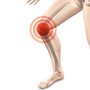
Compression proximal (near) to the elbow

Entrapment occurs proximal (nearer) to the elbow on the lateral (side) position between the brachialis (muscle goes from humerus and inserts to ulna bone of elbow) and brachioradialis (muscle goes from humerus to radius bone at wrist).


 Symptoms/ Signs of Radial Neuropathy

As mentioned previously, there is usually weakness of the triceps if the compression is in the axillary area.  This differentiates higher from lower levels of compression.  Moreover, entrapment from the level of the spiral groove of the humerus down usually does not affect the triceps. 

However, if severe enough, compression in the axilla can be very extensive.  It can lead to loss of elbow, wrist (wrist drop) and finger extension. This is because entrapment at the axilla affects all of the motor and sensory (possible numbness) radial distributions.


Radial Neuropathy/Nerve Entrapment Causes

Firstly, your pain and paresthesias (numbness and tingling) could have come on insidiously (gradually).  However, radial neuropathy/nerve entrapment can be due to various causes such as trauma, tumor, scaring/fascial restrictions, fracture, and tourniquet or crutch use causing pressure at the axilla.

Secondly, Saturday night palsy; where you are sleeping with the head on an outstretched hyperabducted (stretched above head) arm may lead to it as well.

Thirdly, sports such as gymnastics and wrestling may be a cause due to some of the maneuvers.

Fourthly, other sports such as baseball and weight lifting where your elbow is forcefully extended may be a cause.

Fifthly, even runners are at risk for radial neuropathy.  Runner’s Radial nerve palsy occurs due to keeping the elbow tightly flexed while running.  This causes numbness in the forearm and dorsal (upper) hand.

Sixthly, repetitive or prolonged muscle contracture in laborers and throwers may be a cause.


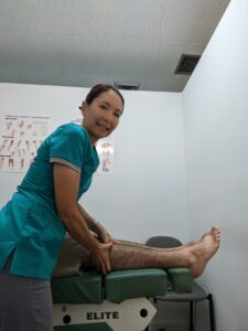
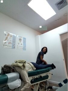
Treatment at Meiri Chiropractic for Radial Neuropathy:

-Soft tissue techniques will be rendered to the contracted fibrotic muscle and collagen at entrapment sites.

-Chiropractic adjustments (chiropractic manipulative therapy) to the shoulder, elbow and associated restricted joints along the kinetic chain and spine is important.

-Therapeutic exercise will be given for stretching and strengthening.

-If trauma or swelling is involved, ice, compression, elevation, and physical therapy modalities is utilized to reduce swelling. Ergonomic recommendations to change the inciting activity is necessary.

Are your suffering from shoulder or arm pain from Radial Neuropathy?

We offer excellent Chiropractic care for Radial Neuropathy. At Meiri Chiropractic, we spend the time necessary to examine, diagnose and treat every neuromusculoskeletal condition and various ailments you have. Indeed, Chiropractic is a holistic and natural way to not only treat shoulder and arm pain, but to keep your body in its best working condition. We have been offering effective chiropractic care in West Palm Beach since 2006. Many of our patients reviews note our excellence. Call us today at 561-253-8984 to make an appointment or to find out more about Shoulder and Arm Pain from Radial Neuropathy: Chiropractic Can Help.


Do you have pain and tenderness around your patellar tendon or lower part of the patella (kneecap)? It may be described as a burning pain in the knee with use or after prolonged sitting with the knee flexed. Perhaps jumping, running or walking may be difficult due to the pain?  Additionally, you may have swelling and erythema (redness of the skin) along the patellar tendon depending on the amount of inflammation.  Also, in chronic cases a nodule of scar tissue that is often tender can be found in the tendon or near the patella.  Keep reading to find out more about Knee Pain Due to Patellar Tendinitis: Chiropractic Can Help.

Knee Anatomy: Femur (thigh bone), Tibia (shin bone), Fibula (thin, outer long bone of the leg), and the Patella (kneecap)

There are three “articulations” (joint or juncture between bones or cartilages) at the knee: the tibiofemoral, patellofemoral, and tibiofibular articulations. However, only the tibiofemoral and patellofemoral articulations participate in knee joint activity. The tibiofibular articulation/joint does not actually contribute to the actions of the knee. Instead, it is part of the ankle joint complex moving the ankle in all the various ranges of motion. However, dysfunctional processes in the proximal tibiofibular articulation/joint can affect other knee functions and can be a source of knee pain.

So the tibiofemoral and tibiofibular are synovial articulations.  These are freely movable joints. And the bony surfaces are coated with hyaline cartilage.  It is unified by a fibrous articular capsule. However, the third articulation, patellofemoral, is a functional joint (non-synovial joint).   


What is Patellar Tendinitis (Jumper’s Knee)?

Tendinitis is inflammation of the thick fibrous cords that attach muscle to bone. Jumper’s knee is a traction (pulling/microtear of knee) overuse syndrome (group of signs and symptoms). Furthermore, it can be a quadriceps (group of muscles in the front of thigh) tendinitis or patellar tendinitis.  So this depends on whether the quadriceps bone-tendon junction of the superior (upper) patella or the bone-tendon junction of the inferior (lower) patella is primarily involved. 

Quadriceps tendinitis is more common in the age group older than 30 years.  And patella tendinitis is more common in the adolescent (10-17 years old) population.  Importantly, involvement of the quadriceps in an adolescent is probably an avulsion (tearing away) of the proximal (nearer to the trunk) patellar epiphysis (growth plate) rather than an actual quadriceps tendinitis. 


Cause of Patellar Tendinitis


Patellar Tendonitis is most often an overuse/ repetitive injury problem.  Most of it is due to “microtearing”.  Next, your body will try to repair the microtearing degenerative process that is taking place. Ultimately, this leads to increased thickness of the insertional fibrocartilage at the patella bone-tendon junction.  Sadly, patients treated with steroid injections are prone to developing ruptures. In contrast, Chiropractic restores and normalizes function to joints.  Therefore, the inflammation and pain subside.

Sometimes, a subtle lateral (outward) subluxation may be a case due to overloading of the patellar tendon.  Moreover, tight hamstrings can produce abnormal movements when extending the knee.  Lastly, tight hamstrings and patellar tendinitis were found as a cause in hard playing surfaces combined with overtraining.


Dr. Natalie Meiri adjusts a patient's knee
Dr. Natalie Meiri adjusts a patient’s knee
 
4 Stages of Patellar Tendinitis

1st Stage-  pain only after activity.

2nd Stage- pain at the beginning of activity that disappears after warm up and returns after activity. 

3rd Stage- constant pain at rest and inability to participate in sports

4th Stage- complete rupture of the patellar tendon.

Patellar Tendonitis is not self limiting (goes away on own).  Without proper treatment and with continued overuse, it may progress through the different stages described above.  Even Stage 3 may sometimes be irreversible and require surgery.  Therefore, it is wise to get treatment as soon as possible.


Treatment at Meiri Chiropractic for Patellar Tendinitis

Firstly, during the acute phase, rest/ proper warm up before use, stretching, ice after use and a patella restraining brace are recommended.

Secondly, soft tissue techniques (e.g. myofascial release, cross friction) will be performed on the patella ligament and all associated muscles. 

Thirdly, chiropractic adjustments (chiropractic manipulative therapy) to the Knee, patella  and associated restricted joints along the kinetic chain is important.

Fourthly, therapeutic exercise will be given for stretching and strengthening.  For example, hamstring and gastrocnemius and quadriceps stretching and strengthening.


Are your suffering from Knee or patellar tendon Pain?


We offer excellent Chiropractic care for Patellar Tendinitis. At Meiri Chiropractic, we spend the time necessary to examine, diagnose and treat every neuromusculoskeletal condition and various ailments you have. Indeed. chiropractic will to keep your body in its best working condition. We have been offering effective chiropractic care in West Palm Beach since 2006. Many of our patients reviews note our excellence. Call us today at 561-253-8984 to make an appointment or to find out more about Knee Pain Due to Patellar Tendinitis: Chiropractic Can Help.


Dr. Natalie Meiri adjusts the patient's foot
Dr. Natalie Meiri adjusts the patient’s foot

Do you have persistent pain, burning, aching, or numbness in the back side of your leg,  ankle, or foot?  These are all symptoms of Sural Neuropathy. Read on to learn about Ankle and Foot Pain Due to Sural Neuropathy: Chiropractic Can Help.


Anatomy of the Sural Nerve

The sural nerve is a cutaneous (affecting the skin) nerve. So it is purely sensory. It provides only sensation to the posterolateral (back outside) aspect of the distal (away from body) third of the leg and the lateral (outside) aspect of the foot, heel, and ankle.

It is formed by terminal (ending) branches of the tibial and common peroneal nerves (both branches of the sciatic nerve) that join together in the distal third of the leg.

The Sural Nerve descends between the two heads of the gastrocnemius (calf muscle).  Next, it pierces the deep fascia (casing of connective tissue holding muscles) in the middle or upper part of the back of the leg. Then it is joined by the sural communicating branch of the common peroneal nerve. Finally, it continues to the lateral malleolus (bump of bone on the outside of your ankle). 

Dr Natalie Meiri adjusts a patient's ankle

Entrapment Sites

Sural nerve entrapment/neuropathy most commonly occurs in the ankle/foot area. The entrapment site located approximately two thirds the distance between the knee and the heel is a less common entrapment location.  The  most common sites are in the fascia just above the ankle and behind the lateral malleolus.


Sural Neuropathy/Nerve Entrapment Causes

Your pain and paresthesias (numbness and tingling) could have come on insidiously (gradually).  However, Sural neuropathy/nerve entrapment can be due to various causes such as trauma, bakers cyst, scaring, fascial restrictions and vascular problems.

Firstly, tight boots and lacing may be the cause of compression of the sural nerve. 

Secondly, different forms of trauma can be a leading cause of sural nerve pain. For instance after an ankle sprain or an ankle fracture.  Afterwards, this can create significant pulling and traction on the nerve. Additionally, direct impact to the nerve from being kicked, falling, or an accident can be a cause.

Thirdly, entrapment sites are areas along the nerve’s pathway that may become entrapped, causing more pressure on the nerve. For instance, like carpal tunnel syndrome in the hand.

Fourthly, trauma from surgery can cause sural neuropathy. Sadly, this may be a common cause of chronic pain CRPS (Complex regional pain syndrome). And this is often in the news because of the American opiate crisis.   Patients are taking harmful drugs due to pain. And some of these patients’ pain could be related to this painful sural neuropathy. Various surgeries in the ankle, foot and leg can injure the sural nerve, resulting in sural nerve pain.

 
Here are some examples of  Sural Neuropathy after surgery:
  • Heel Fractures: Surgey is performed on the outer side of the heel bone which is next to the pathway of the nerve. A post-surgical scar can occur which irritates the nerve. So the nerve can become entrapped and tangled in the scar.
  • Achilles Tendon Repair: Similarly, surgery performed on the achilles tendon for repair or lengthening places the nerve at a risk for injury.
  • Ankle Surgery: Any ankle surgery may also cause injury to the nerve.
Dr. Natalie Meiri adjusts a patient's knee/leg
Dr. Natalie Meiri adjusts a patient’s knee/leg
Treatment at Meiri Chiropractic for Sural Neuropathy

First, soft tissue techniques will be rendered to the contracted fibrotic muscle and collagen at entrapment sites.

Second, chiropractic adjustments (chiropractic manipulative therapy) to the foot, ankle and associated restricted joints along the kinetic chain is important.

Third, therapeutic exercise will be given for stretching and strengthening.

Lastly, if trauma or swelling is involved, ice, compression, elevation, and physical therapy modalities is utilized to reduce swelling. Also, an orthotic and/or correction of shoes may be necessary. 

Are your suffering from leg, ankle or foot pain from Sural Neuropathy?

We offer excellent Chiropractic care for Sural Neuropathy. At Meiri Chiropractic, we spend the time necessary to examine, diagnose and treat every neuromusculoskeletal condition and various ailments you have. Indeed, Chiropractic is a holistic and natural way to not only treat leg, foot and ankle pain, but to keep your body in its best working condition. We have been offering effective chiropractic care in West Palm Beach since 2006. Many of our patients reviews note our excellence. Call us today at 561-253-8984 to make an appointment or to find out more about Ankle and Foot Pain from Sural Neuropathy: Chiropractic Can Help.


If you have knee pain from Pes Anserinus Bursitis, Chiropractic can provide relief! This post is about Knee Bursitis and Chiropractic Treatment: Pes Anserinus Bursitis.


What is bursitis?

Bursae are fluid-filled, sac like cavities. Bursae are located where muscles and tendons move over bony joint areas. They reduce friction caused by muscles and tendons moving against skin and bones.  Also, they facilitate movement.

A knee bursa is like a cushion. So when one becomes inflamed, increased tension and pain can occur in a condition known as bursitis. The bursa in the knee increases with fluid and redness in the area it occurs.


Where is the Pes Anserinus and the bursa?

The pes anserinus is composed of the combination of tendinous insertions of the sartorius, gracilis, and semitendinosus muscles:


  1. sartorius– long, thin, superficial muscle that runs down the length of the thigh in the anterior (front) compartment
  2. gracilis– muscle descends almost vertically down the leg and inserts on the medial tibia at the Pes anserinus
  3. semitendinosus– is one of the three muscles that make up the hamstrings muscle group, and it is located at the posterior (back) and medial (inner) aspect of the thigh.

The pes anserinus bursa lies about 2 inches below the medial knee joint line between the pes anserinus (bracilis, sartorius, and semitendinousus muscles) above and the medial collateral ligament and anterior tibia (front of shinbone) below. 


Causes of Pes Anserinus Bursitis

Firstly, one possible cause is over training such as excessive miles, excessive hills, and improper or inadequate stretching.  This can lead to shortened medial hamstrings which may compress the bursa with overuse.

Secondly, knee valgus (Knock knee) and heel valgus (heel to drift outwards) may also lead to pes anserinus bursitis.

Thirdly, external tibial torsion (leg turns outward), femoral anteversion (inward twisting of the thigh bone) and a broad pelvis (in women) can cause pes anserinus bursitis.

Fourthly, if you have knee osteoarthritis, there’s a 75% chance of also having pes anserinus bursitis.

Fifthly, it occurs more often in 50-80 year old women especially if overweight. 

Finally, it is also found in runners and soccer players.


Symptoms and Signs of a Pes Anserinus Bursitis

You may have pain that occurs with walking up stairs.  This may occur in the early stages of training or later stages of use (distance running).  Furthermore, you usually will have pinpoint tenderness 2 inches below the medial (inner) knee joint line.  And there may be moderate swelling.  Also, your pain may radiate into the medial (inner) knee joint and along the medial (inner) hamstring.  


Dr. Natalie Meiri adjusts a patient's knee
Dr. Natalie Meiri adjusts a patient’s knee


Treatment for Pes Anserinus Bursitis at Meiri Chiropractic

First, biomechanical evaluation of the lower extremity regarding torsion and angulation is necessary. 

Second, chiropractic manipulative therapy (adjustments) is rendered to the knee and associated spine and extremity (limb) joints. 

Third, manipulation of muscle contractures with soft tissue techniques is utilized.  Some Techniques include gentle PNF/PIR (e.g. post isometric relaxation) and/or myofascial release techniques.  

Finally, therapeutic exercises for rehabilitation/ strengthening are prescribed.  Flexibility and strengthening of the hamstrings, gastrocnemius and soleus are important.  And corrective shoes or orthotics for heel and knee valgus maybe necessary.


Getting regular chiropractic treatment at a chiropractic center in West Palm Beach can help naturally relieve the pain and dysfunction of the knee. Chiropractic is a holistic and natural way to not only treat existing conditions, but to keep your body in its best working condition.

Contact Meiri Chiropractic today at 561-253-8984 to make an appointment or to learn more about  Knee Bursitis and Chiropractic Treatment: Pes Anserinus Bursitis.


If you have knee pain from a bakers cyst, Chiropractic can provide relief! This post is about Knee Bursitis and Chiropractic Treatment: Baker’s Cyst.


What is bursitis?

Bursae are fluid-filled, sac like cavities. Bursae are located where muscles and tendons move over bony joint areas. They reduce friction caused by muscles and tendons moving against skin and bones.  Also, they facilitate movement.

A knee bursa is like a cushion. So when one becomes inflamed, increased tension and pain can occur in a condition known as bursitis. The bursa in the knee increases with fluid and redness in the area occurs.


Symptoms and Signs of a Baker’s Cyst

You may have swelling, aching and tenderness in the popliteal area of the knee.   The sac is located in the medial (inner) side of the popliteal fossa (diamond-shaped space behind the knee joint).


The Baker’s Cyst

The baker’s cyst is an idiopathic (cause unknown) form of bursitis where the cyst contents are not in the knee joint.

It can be found in children between ages 6-8 and 12-14 following rapid growth. Baker’s cysts in children are painless and normally disappear as the child matures. 

In contrast, a baker’s cyst found in adults is symptomatic. In adults it has intra-articular (within joint) problems such as degeneration of posterior horn of the medial meniscus (C-shaped cartilage between shin bone and thigh bone) or synovial membrane (connective tissue joint lining) irritation. 

Apparently, a baker’s cyst is due to swelling of the semimembranosus bursa.  The semimembranosus bursa is located between the semimembranosus (one of a group of muscles called the Hamstrings) and gastrocnemius (calf muscle) tendon.  Therefore, it is also called semimembranosus bursitis. Interestingly, poor flexibility of the semimembranosus and gastrocnemius muscles could contribute to development of a baker’s cyst. And this results in tendinitis.

Furthermore, degenerative joint disease or a torn meniscus are other possible contributing causes for a baker’s cyst.


Treatment for a Baker’s Cyst at Meiri Chiropractic

First, Chiropractic care for the knee conditions (e.g. tendinitis) can resolve the cyst. 


Second, chiropractic manipulative therapy (adjustments) is rendered to the knee and associated spine and extremity (limb) joints.


Third, manipulation of muscle contractures with soft tissue techniques is utilized.  Some Techniques include gentle PNF/PIR (e.g. post isometric relaxation) and/or myofascial release techniques. 


Finally, therapeutic exercises for rehabilitation/ strengthening are prescribed. Especially, the lack of flexibility in the hamstrings and gastrocnemius are addressed.


Getting regular chiropractic treatment at a chiropractic center in West Palm Beach can help naturally relieve the pain and dysfunction of the knee. Chiropractic is a holistic and natural way to not only treat existing conditions, but to keep your body in its best working condition.

Contact Meiri Chiropractic today at 561-253-8984 to make an appointment or to learn more about  Knee Bursitis and Chiropractic Treatment: Baker’s Cyst.


i


Do you have numbness and tingling across the bottom of the foot?  Furthermore, do you have burning pain in the ankle or foot?  It may be often at the sole or first toe. Your pain and paresthesias (numbness a tingling) probably came on insidiously (gradually).  And you do not have any associated low back or leg pain. You may also have noticed weakness in your foot. Lastly, your symptoms are worse by prolonged standing, walking, or running.  These are all symptoms of Tarsal Tunnel Syndrome.

In one study that evaluated patients with tarsal tunnel syndrome, 45 % reported numbness, 39% reported pain, and 16% reported both.  Read on to learn about Ankle and Foot Pain from Tarsal Tunnel Syndrome: Chiropractic Can Help.

Cause

The posterior tibial nerve may be stretched or compressed in the tarsal tunnel.  This is a tunnel formed by the flexor retinaculum (band of thickened deep fascia around tendons), calcaneus (large bone forming the heel), distal tibia (shin bone), and malleolus (inner and outer boney protuberances of ankle).

Contents of the tunnel include: the tendons of the posterior tibialis (muscle in posterior compartment of the lower leg), flexor digitorum longus (also located in posterior compartment muscles of lower leg), and flexor hallucis (muscle goes from calf to 1st toe) and the posterior tibial artery, vein, and nerve. 

Anatomy of the Tibial Nerve

Firstly, the tibial nerve is one of two main terminal branches of the sciatic nerve.  And the sciatic nerve is the largest nerve in the human body.

Secondly, the tibial nerve originates from the L4-S3 spinal nerve roots and provides motor and sensory innervation to most of the posterior (back part) leg and foot.

Thirdly, the tibial nerve descends from the popliteal fossa (space behind knee joint) into the posterior compartment of the leg. It passes deep to the fibular and tibial heads of soleus muscle (a calf muscle found between knee and heel). 

Fourthly, it runs vertically through the deep layer of the posterior (back) compartment of the leg, along with the posterior tibial blood vessels. The tibial nerve provides innervation to the surrounding muscles in this area.

Fifthly, the tibial nerve exits next at the ankle joint. It passes behind the medial malleolus (boney protuberance at inner ankle) to enter the sole of the foot.  Here it supplies innervation to most of the intrinsic (deep) muscles and skin.

Sixthly, past the tarsal tunnel, the tibial nerve gives off its terminal branches, the medial and lateral plantar nerves.  These nerves continue to supply motor and sensory innervation to the plantar foot.


Pronation of the Foot

Hyperpronation (excessive inward rotation of the foot during gait) is often the cause of tarsal tunnel syndrome.  This could be due to tightening of the flexor retinaculum or arch of the abductor hallucis muscle. This muscle is located on the inner border of the foot and bends the big toe and moves it out to the side. However, trauma and the subsequent swelling may also cause entrapment of the tibial nerve. 

There may also be a chronic tenosynovitis of the posterior tibial or flexor digitorum longus tendons.  This occurs near the malleolus (prominent bones at ankle) as areas that can compress the tibial nerve.  These fascial restrictions may be a primary cause. However, it is necessary to rule out ganglions, myositis ossificans, exercise compartment syndrome, and tumors.


Examination

Your doctor may order a nerve conduction study in a difficult unremitting case.  However, usually patients are diagnosed by the physical (orthopedic, neurological etc.) examination.


Dr Natalie Meiri adjusts a patient's ankle
Dr Natalie Meiri adjusts a patient’s ankle


Treatment at Meiri Chiropractic for Tarsal Tunnel Syndrome

First, soft tissue techniques will be rendered to the contracted fibrotic muscle and collagen at entrapment sites. Second, chiropractic adjustments (chiropractic manipulative therapy) to the foot, ankle and associated restricted joints along the kinetic chain is important.

Third, Therapeutic exercise will be given for stretching and strengthening.

Lastly, if trauma or swelling is involved, ice, compression, elevation, and physical therapy modalities is utilized to reduce swelling. Also, an orthotic and/or correction of shoes may be necessary if there is pronation of the foot.


Are your suffering from ankle or foot pain from Tarsal Tunnel Syndrome?

We offer excellent Chiropractic care for Tarsal Tunnel Syndrome. At Meiri Chiropractic, we spend the time necessary to examine, diagnose and treat every neuromusculoskeletal condition and various ailments you have. Indeed, Chiropractic is a holistic and natural way to not only treat foot and ankle pain, but to keep your body in its best working condition. We have been offering effective chiropractic care in West Palm Beach since 2006. Many of our patients reviews note our excellence. Call us today at 561-253-8984 to make an appointment or to find out more about Ankle and Foot Pain from Tarsal Tunnel Syndrome: Chiropractic Can Help.


Meiri Chiropractic

5601 Corporate Way, Ste#102

West Palm Beach, FL 33407